Video Details
|
Apical Dentinal Cracks as a Cause of Endodontic Treatment Failure: Microsurgical Diagnosis and Management part 1 of 2
Description:
Non surgical endodontic treatment is a predictable and reliable treatment with high success rates ranging from 86%–98% . Nevertheless, for a variety of reasons, endodontic failure still occurs, and presence of clinical signs and symptoms along with radiographic evidence of periapical bone destruction indicates the need for retreatment.
The first and most important step for retreatment planning is to determine the cause of endodontic failure.
The failure of the root canal treatment is generally attributed to either residual or resistant intraradicular microorganisms surviving cleaning and shaping procedures or new microorganisms invading the canals via miclroleakage. Rarely some persistent lesions may be attributed to extraradicular infection, foreign body reaction or a true periapical cyst. Moreover, the outcome of root canal retreatment is less favorable than the initial treatment.
Endodontic failure related to microorganisms can be caused by procedural errors such as root perforation, ledge formation, separated instruments, missed canals, as well as anatomical difficulties such as apical ramification, isthmuses, and other morphologic irregularities. Nevertheless, a precise diagnosis can be made only after surgery or extraction. A precise inspection of the root apex or resected root surface is one of the best advantages of endodontic microsurgery. It helps identify the cause of endodontic failure, so that causative factors can be removed completely during the surgical procedure.
This new lecture is about analyzing the cause of my personal retreatment failures by using microsurgical techniques. It seems that apical dentinal cracks dominate my failures.
Preventive measures, risk factors and treatment options are also discussed.
Date Added:
5/14/2012
Author(s):
Antonis Chaniotis, DDS MDSC
Chaniotis Antonis graduated from the University of Athens Dental School (Greece) in 1998. In 2003 he completed the three-year postgraduate program in Endodontics at the Univer...
[read more]
|

|
Online Videos / General / Endodontics / Apical Dentinal Cracks as a Cause of Endodontic Treatment Failure: Microsurgical Diagnosis and Management part 1 of 2
Questions & Comments
|
Jasper Ainslie - (1/17/2018 6:38 PM)
thankyou
|
Robert Burstein - (12/23/2014 9:18 AM)
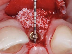
Any thoughts of management of the boney void with graft material and/or membrane to avoid ingress of epithelial tissue? Great lecture and video thank you.
|
NAGARAJAN MUTHUSAMY - (3/12/2014 4:37 AM)
Great presentation on peri- apical surgery.very nice.
|
Maurice Salama - (10/23/2012 7:05 PM)
Clean surgery and great endo. Applause.
Dr. Salama
|
Dimitris Tsanaktsidis - (7/12/2012 10:38 AM)
5* from me . Especially for the Greek Version. Well done!
|
milind saudagar - (6/30/2012 1:33 PM)
indeed very impressive. with the era of fast rotary and greater taper endodontics, cracks in root are becoming a common feature. we tend to please ourselves radiographically with big huge fillings ignoring the anatomy of the cannal and thus compromising the tooth structure and strength. very well and neat sealing of the isthmus. only a master can do it. thanks
|
Antonis Chaniotis - (5/20/2012 3:53 AM)
Thank you dr. Scholl. I place a pellet in the bone crypt and leave it there until the surgery is finished. High concentration epinephrine anesthetics, pressure hemostasis and epinephrine pellets most of the times would be sufficient. Be carefully with the racellets in a maxillary sinus communication. You can have sterile saline if your dental unit have a Tank for delivering sterile saline. Most of the times the impact air 45 handpiece is used with water . You can perform the final rinse with sterile saline, or use a surgical motor for the apicoecTomy. Piezo surgery units are also an excellent choice!
|
Paul Scholl - (5/19/2012 8:06 PM)
Nice case. What is the protocol for hemostasis with the pellets? How do you set up your surgical high speed to flow sterile fluid onto the site? Thanks for the informative video
|
Antonis Chaniotis - (5/19/2012 10:59 AM)
Thank you doctor. Preventive measures and risk factors follow in part II.
|
Related Videos |
|
|
|
Three-Canal Maxillary Premolar Teeth: A Clinical Reality
To treat three-canal maxillary premolar teeth predictably, it is necessary to be aware of the clinical and radiographic anatomy. These teeth may also require special shaping and obturation techniques.
The purpose of this presentation is to show the complete procedure of an endodontic treatment of a upper left second premolar with three root canals.
Presented By:: |
Carlos Alfonso Morais, DDS |
Presentation Style: |
Video |
Community Rating: |
|
|
Watch Now>>
|
|
|
|
|
Prevention and Management of Broken Endodontic Instruments
he separation of endodontic instruments is a procedural accident that results in major treatment challenges and medico legal considerations in endodontics. With the advent of rotary nickel-titanium (Ni Ti) instrumentation the issue became extremely prominent. Extensive research has been conducted to understand the mechanics of instrument separation. This has led to changes in the manufacturing methods, instrument design and instrumentation protocols. Most recently, reciprocation is challenging rotary instrumentation in terms of speed and safety from fracture. Although the incidence of file breakage can be drastically decreased, it cannot yet be eliminated. No matter what instruments you are using inside the root canal system, the prevention and management of broken endodontic instruments still remains a hot topic.
Presented By:: |
Antonis Chaniotis, DDS MDSC |
Presentation Style: |
Video |
Community Rating: |
|
|
Watch Now>>
|
|
|
|
|
Prevention and Management of Broken Endodontic Instruments
This lecture aims to review the most common preventive measures to avoid file breakage and to provide clinical tips and recommendations for the management of challenging broken file cases. Highly invasive, minimal invasive and non invasive broken instrument removal techniques are showcased through video case presentation series under the microscope.
Presented By:: |
Antonis Chaniotis, DDS MDSC |
Presentation Style: |
Video |
Community Rating: |
|
|
Watch Now>>
|
|
|
|
Related Courses |
|
|
Prevention and Management of Broken Endodontic Instruments
This lecture aims to review the most common preventive measures to avoid file breakage and to provide clinical tips and recommendations for the management of challenging broken file cases. Highly invasive, minimal invasive and non invasive broken instrument removal techniques are showcased through video case presentation series under the microscope.
Presented By:: |
Antonis Chaniotis, DDS MDSC |
Presentation Style: |
Online Self-Study Course |
CE Hours: |
1 CEU (Continuing Education Unit) |
|
Watch Now>>
|
|
|
|
|
Optimized Instrumentation & Technology in Endodontic Therapy
Rotary instrumentation using Optimized Torque Reduction (OTR) motion and Apex Locator-Handpiece technology has the potential to improve clinical efficiency during endodontic therapy. In this webinar, Dr. Nasseh explains the implementation of one such technology using the ESX instrumentation and obturation system. He further shares some tips and settings for effective, safe operations. Various endodontic motions and their indications for use are explained and an anterior tooth demonstration is presented to consolidate all covered points with a clinical demonstration.
Presented By:: |
Allen Ali Nasseh, DDS, MMSc |
Presentation Style: |
Online Self-Study Course |
CE Hours: |
1 CEU (Continuing Education Credit) |
|
Watch Now>>
|
|
|
|
|
CBCT in Endodontics: Changing the Landscape of Diagnosis and Clinical Treatment
Radiographic imaging is essential in the diagnosis, treatment planning and follow-up in endodontics. The interpretation of an image can be confounded by a number of factors, including the regional anatomy as well as superimposition of both the teeth and surrounding dento-alveolar structures. As a result of superimposition, periapical radiographs reveal only limited aspects, a 2-dimensional view, of the true three dimensional anatomy. Additionally, there is often geometric distortion of the anatomical structures being imaged with conventional radiographic methods. These problems can be overcome by utilizing small or limited volume cone beam computed tomography (CBCT) imaging techniques which produce accurate 3-dimensional images of the teeth and surrounding dento-alveolar structures. This presentation will highlight the indications, advantages and considerations of the use of CBCT in endodontics.
Presented By:: |
Frederic Barnett, DMD |
Presentation Style: |
Online Self-Study Course |
CE Hours: |
1 CEU (Continuing Education Unit) |
|
Watch Now>>
|
|
|
|
Related Articles |
|
|
|
Dental Endoscopic Viewing System
Endoscopy has enabled medicine to take a quantum leap in disease diagnosis and therapeutic management by allowing visualization and access for repair of injuries via minimally invasive surgical (MIS) techniques. Since their introduction, fiberoptic bundles and electronic video technology have continually advanced and fostered smaller-bore (viewing tubes) endoscopes of 2 mm and less, further improving access into smaller cavities and anatomical spaces of the body. This viewing technology has reached…
Author(s): |
Ronald Goldstein, DDS;David Garber, DMD;Maurice Salama, DMD;Henry Salama, DMD;Lee H. Silverstein, DDS, MS, FACD, FICD |
|
View Article>>
|
|
|
|
|
|