Video Details
|
Autogenous Bone Graft - Part 3: Contouring & Fixation
Description:
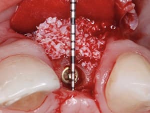
In part 3 of this series, Dr. Michael Pikos demonstrates the techniques for contouring and fixation of a cortical autogenous graft harvested from the ramus buccal shelf. In addition, specific techniques and guidelines are suggested in ensuring tension free closure of the flap over the graft. Membrane use and PRP are also discussed
Date Added:
4/6/2009
Author(s):
|

|
Online Videos / Surgery / Bone Grafting / Autogenous Bone Graft - Part 3: Contouring & Fixation
Questions & Comments
|
Maurice Salama - (2/18/2015 9:29 PM)
I prefer doing the periosteal realeasing incision just prior to wound closure and final suturing when possible as it allows me to control graft positioning, contouring and membrane placement PRN as well as final stabilization of all of the above without having to manage bleeding and visibility at the same time.
|
michael abba - (1/22/2013 8:58 AM)
Dr korner,
I agree about the periosteal incision , but I think it was done because of "filming purposes"
|
Robert Körner - (1/21/2013 1:38 PM)
Is there nobody out there who asks himself: isn´t this a case that could (or should) have been treated without augmentation?(or at the outmost a little lateral augmentation in the most medial area)
Three Implants of at least 10mm length and sufficent diameter shouldn´t have been a problem.By the way - where are the guidelines for the "crown-implant ratio".Actually we do not really know much about the apropriate legth and diameter of the implants we use. Further I think that is prefareable to do the periosteal incision much more prior to woundclosure, because bleeding will have stopped,when you start suturing, and you have less risk of haematoma.
|
Michael Pikos - (4/20/2010 11:29 AM)
Hi guys,
Sorry I haven’t been more punctual with regard to being part of this blog. Allow me to address some of the questions and concerns that have been posted already regarding flap management for the RBS bone harvest. First, generally speaking, contouring a block graft prior to fixation is always indicated. There are times however where post fixation contouring still needs to be done and when this occurs it can be done predictably with a slow speed burnishing concept that I have shown. Yes, this can put the block at risk but not if it’s done properly. I do utilize particulate mineralized allograft (MinerOss) for any morticing that is necessary with these block grafts and especially when overcontouring is indicated. For the past 4 years now I utilize PRGF as opposed to PRP as a bioactive modifier with all of my bone grafting including sinus grafting from autogenous block grafting, ridge splitting, etc.
The importance of flap relaxation prior to closure of any bone graft be it block bone, mesh particulate, ridge split, etc. cannot be emphasized enough. This technique that I show is of course specific to the mental neurovascular bundle area which not too coincidentally happens to be the area (posterior mandible) where most bone grafts fail – due to incision line opening and / or vestibular dehiscence. More often than not the clinician is too conservative with flap relaxation in and around the neurovascular bundle. Hopefully this helps. Also, it is never a good idea to place implants simultaneously with block grafting in that the remodeling that will occur will result in graft compromise as Dr. Salama pointed out. Always best to stage implant placement with block grafting. Again, hope this helps clarify some points.
Best regards,
Dr. Pikos
|
Fadi Assaf - (4/19/2010 8:40 AM)
Would it be safer to use an electric motor?
|
Maurice Salama - (1/4/2010 12:10 PM)
Milind and Mohammed; Thanks for the posts and questions. I do not believe leaving behind periosteum would have a negative effect on the healing but leaving soft tissue of any kind would be a negative. Denuding the bone as Dr. Pikos shows increases the bleeding of the recepient site allowing mesenchymal cells to emerge from the host bone allowing for better incorporation of the grafted bone. As for placing implants simultaneously, I would avoid that as it increases the risk for a large failure of the grafted area. Better to graft and then come back 4-6 months later and place the implants when dealing with any kind of block graft autogenous, allograft or xenograft. As an additional comment, the biggest risk with these blocks is early flap dehiscence or fenestration. Always make sure to work in a thick tissue environment and gain tension free closure. Hope this helps and welcome to the blog Dr. Salama
|
mohammed shakeel - (1/3/2010 10:34 PM)
could u have placed implants at the same time??
|
milind saudagar - (1/3/2010 7:11 AM)
just watched the surgery. it was very good procedure. my doubt was the bone part which was fixed appeared to be completely denuded of all periosteum & blood. if some amount of tissue would have been present, will it affect healing in any manner?
|
Rand Ollerton - (4/26/2009 4:30 PM)
Very nice description of buccal release in such a way as to not violate the inferior alveolar nerve.
|
|
Related Videos |
|
|
|
|
|
|
Alveolar Ridge Regenerative Strategies: Autogenous Bone vs BMP-2
This clinical based presentation will compare the use of autogenous bone vs BMP-2 for alveolar ridge reconstruction. The science, indications, advantages and disadvantages of each approach will be featured. Single tooth to full arch reconstruction cases will also be shown along with understanding the application of non-resorbable vs resorbable mesh barriers for alveolar ridge reconstruction.
Presented By:: |
|
Presentation Style: |
Video |
Community Rating: |
|
|
Watch Now>>
|
|
|
|
|
Bone Graft Cements: A New Advance to Augmentation Solutions
The purpose of this lecture is to shed light on the evolution of raw materials in order to find the ultimate bone graft cement, exploring the potential and the new opportunities of using cement-based augmentation materials in the maxillofacial and dental fields, and emphasizing the advantages, disadvantages and methods of use from the scientific and clinical points of view.
Presented By:: |
Amos Yahav, DMD |
Presentation Style: |
Video |
Community Rating: |
|
|
Watch Now>>
|
|
|
|
|
Subperiosteal Implants - The New Approach for the Extreme Atrophic Jaws
Subperiosteal implants started in the 40’s but were discontinued due to the poor success rates in the long term.
Today, 80 years later, a completely different technology has emerged, bringing Subperiosteal implants to the next level.
Presented By:: |
Bernardo Nunes de Sousa, DDS, MSc |
Presentation Style: |
Video |
Community Rating: |
|
|
Watch Now>>
|
|
|
|
Related Courses |
|
|
Complete Management of the Implant Site: From Simple Cases to Complex Situations
The health and the structure of the peri-implant tissue is vital for a long term prognosis of aesthetic and functional dental treatments. The prevalence of peri-implant diseases and recessions is a problem that must be overcome by a perfect treatment planning, along with a step-by-step design of the future restorations. When the clinician places an implant, he must consider the 3D positioning regarding the hard and soft tissue. When one decide to restore an implant, he must be aware of the gingival volume and it’s position. In this presentation, we want to show fully documented cases, from bone grafting, soft tissue enhancement, to final restorations.
Presented By:: |
Mihnea Cafadaru, DDS |
Presentation Style: |
Online Self-Study Course |
CE Hours: |
1 CEU (Continuing Education Unit) |
|
Watch Now>>
|
|
|
|
|
Hard and Soft Tissue Augmentation: Optimizing Esthetic Results for the Restorative Dentist
Contemporary patient expectations have made esthetics a major requisite of all treatment plans, especially in situations where there is a high smile line. Although new restorative materials have highly improved predictability and esthetic outcomes, soft and hard tissue management play a fundamental role when working in esthetic areas. To achieve ideal esthetics, preservation of the natural soft and hard tissue architecture is a primary clinical objective. These new proposed techniques illustrate the importance of pre-prosthetic soft and hard tissue management when working in highly esthetic compromised areas.
Presented By:: |
Giuseppe Cicero, DDS |
Presentation Style: |
Online Self-Study Course |
CE Hours: |
1 CEU (Continuing Education Unit) |
|
Watch Now>>
|
|
|
|
|
Biological Bone Augmentation- Only for REAL BONE BUILDERS
This webinar is addressed to all clinicians, who want to learn safe and predictable bone harvesting and augmentation techniques for different clinical situations. Clear guidelines for the use of augmentative procedures like the Split bone technique by Prof. Khoury and Biological Bone Augmentation with pure autogenous bone and advanced soft tissue management in order to achieve predicable results will be provided.
Presented By:: |
Frank Zastrow, DDS, M.Sc. |
Presentation Style: |
Online Self-Study Course |
CE Hours: |
1 CEU (Continuing Education Unit) |
|
Watch Now>>
|
|
|
|
Related Articles |
|
|
Intraoral Autogenous Block Onlay Bone Grafting for ExtensiveReconstruction of Atrophic Maxillary Alveolar Ridges
Autologous bone grafting used with dental implants was originally described by Bränemarketal in 1975, and is now a well-accepted procedure in oral and maxillofacial rehabilitation.Placement of an endosseous implant requires sufficient bone volume for complete bone coverage. Furthermore, the pattern of ridge resorption contributes to an unfavorable maxillomandibular relationship, requires angulations of the implants and/or angled abutments, and affects the proximity of adjacent facial concavities(maxillary sinus, nasal cavity) and vital structures(mandibular nerve).
|
|
|
|
Guided Bone Regeneration Using a Titanium Membrane at Implant Placement: A Case Report and Literature Discussion
Reconstruction of the oral supporting tissues lost by disease or trauma is essential to tooth replacement with dental
implant therapy. This treatment requires evidence based augmentative procedures combined with up-to-date and
current techniques. Guided bone regeneration (GBR) aims to initialize this process of alveolar ridge reconstruction by
utilizing biologically active and supportive materials best coupled to the body’s healing processes. The use of nonresorbable,
titanium membranes can achieve GBR by ensuring graft stability and space maintenance so as to ensure
optimal neovascularization. Hereafter is a case report of a ridge defect reconstructed at implant placement, with the
rationale and current, evidence-based literature discussed.
Author(s): |
Howard Gluckman, BDS, MChD, PhD;Jonathan Du Toit |
|
View Article>>
|
|
|
|
|
|