Video Details
|
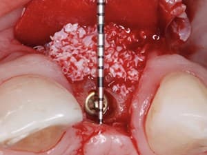
Autogenous Bone Graft - Part 4: Suturing and Tension Free Closure
Description:
In part 4 of this video instructional series of a cortical autogenous graft harvested from the ramus buccal shelf, Dr. Michael Pikos demonstrates the surgical techniques for suturing and tension free closure which are absolutely required for successful osseous augmentation procedures.
Date Added:
4/21/2009
Author(s):
|

|
Online Videos / Surgery / Bone Grafting / Autogenous Bone Graft - Part 4: Suturing and Tension Free Closure
Questions & Comments
|
edward shapiro - (8/26/2014 11:21 PM)
dr. pikos. very nice closure. do you prefer blunt dissection for release of flap in this area? what is the success rate of vertical growth and how much height was regenerated? thanks for the video.
|
yazad gandhi - (2/5/2010 9:34 PM)
Thanks Maurice,
Have taken a biopsy specimen lets see. Will keep you posted. As regards the sterility breakdown that should not be the cause as it was done in the OT under strict asepsis.
Yazad Gandhi
|
Maurice Salama - (2/4/2010 6:25 AM)
Dr. Gandhi; A very rare case failure. Very difficult to assess the reason. Did you take a culture or biopsy of the fibrous tissues and black bone? It would be needed to see what type of bacteria was present. Did the patient have a significant medical history or did the patient have any post surgerical illness? Without this information it would be a guess as to why this may have occured. Without flap breakdown or significant medical issues, I see no reason for the failure other than sterility breakdown. The resorption of the bone, especially the Bio-Oss is almost impossible at 4 1/2 months? Do a oral gingival culture and a blood test on this individual and have an ENT to evaluate the health of their Sinus before performing any other surgical procedures.
Dr. Salama
|
yazad gandhi - (2/4/2010 5:39 AM)
Dr.Pikos and Dr.Maurice,
Would like to know the reason for the failure of a case. Did a Corticocancellous block graft fixation in the maxilla (iliac bone) in a case of Ectodermal Dysplasia. B/L sinus grafts were done using Autogenous & BIOSS 50:50 with biomend extend membrane. No perforations were there in the membrane which was tough n fibrous. 4 n 1/2 mths later the maxilla displayed fibrosis and upon reentry the graft had almost totally disappeared n left behind soft black bone as appears in aseptic necrosis. Almost all of the sinus graft has resorbed whichn has never happened with me till date and the membranes B/L are thinned out and perforated. All this despite there being no infective foci around. Please comment on the possible causes. Thanx
|
Maurice Salama - (8/11/2009 11:47 AM)
Leonardo; Difficult situation. If there is exudate the block is probably not going to do well and should be removed, irrigated and sutured before coming back to regraft at a later date. Would prefer grafting soft tissue prior to 2nd surgery to make sure you have good quality tissue to cover the bone graft. If the block was secure with no exudate, I prefer to grind the exposed bone down until I see a bleeding surface and see if we get epithelialization over the top. Normally, we lose the exposed portion at the least. Good luck Dr. Salama
|
Leonardo Diaz - (8/10/2009 7:37 PM)
would like to know what colud I do with a patient with a deshicence in anterior maxilla after bone grafting.
I sutured tension free with dermis membrane alloderm the right site bone block is ok, but the left site is open with a little exudate after 15 days Post-op.
I take off partially the alloderm because a half was loose and the oter half fixed and sensitive pain.
i need to take a defenitive treatment, so could i do the flap again and a new alloderm leaving the bone block or taking this out.
|
Sean Peng - (4/30/2009 1:04 AM)
Nice and smooth surgery. I’m looking forward to see some videos about complication management during surgery, such as soft tissue bleeding, intra bony bleeding and sinus membrane perforation. I know it’s hard to collect that kind of video, but it makes Dentalxp different from other similar website. I think we should be able to manage complications comfortably before becoming a good surgeon.
|
Related Videos |
|
|
|
|
|
|
Bone Grafting & Implant Dentistry; Classification, Surgical Site Understanding & Membrane Selection Part 2 of 3
Once a tooth is extracted, the natural wound-healing cascade paired with irreversible alteration occurs. Hard tissue grafting techniques combined with the sound understanding of the surgical site theater will help the practitioner to make the proper decision in order to successfully treat their patient. The usage of biological modifiers and surgical barriers are proposed to assist in improving the predictability of the advance surgical care.
Presented By:: |
Alexandre-Amir Aalam, DDS, FICD |
Presentation Style: |
Video |
Community Rating: |
|
|
Watch Now>>
|
|
|
|
|
|
Horizontal Bone Augmentation Using the “SPLIT BONE BLOCK TECHNIQUE” Step by Step - Part 1 of 2
Bone atrophies are one of the most challenging situations in modern implantology. Several techniques have been proposed to be able to place implants in those atrophic situations. This first presentation will show step by step the Split Bone block technique as described by Dr. Khoury to perform horizontal bone augmentation procedures. Bone biology is also explain in order to understand why this technique offer us some great advantages.
The second presentation will focus of vertical bone augmentation procedures using this same technique but through a tunnel approach. The advantages of this approach will also be described in this presentation.
Presented By:: |
Jose L. Dominuez-Mompell, DDS, MsC;Juan Lara Chao, DDS, MsC |
Presentation Style: |
Video |
Community Rating: |
|
|
Watch Now>>
|
|
|
|
Related Courses |
|
|
|
Hard and Soft Tissue Augmentation: Optimizing Esthetic Results for the Restorative Dentist
Contemporary patient expectations have made esthetics a major requisite of all treatment plans, especially in situations where there is a high smile line. Although new restorative materials have highly improved predictability and esthetic outcomes, soft and hard tissue management play a fundamental role when working in esthetic areas. To achieve ideal esthetics, preservation of the natural soft and hard tissue architecture is a primary clinical objective. These new proposed techniques illustrate the importance of pre-prosthetic soft and hard tissue management when working in highly esthetic compromised areas.
Presented By:: |
Giuseppe Cicero, DDS |
Presentation Style: |
Online Self-Study Course |
CE Hours: |
1 CEU (Continuing Education Unit) |
|
Watch Now>>
|
|
|
|
|
Related Articles |
|
|
|
Modified IVAN Technique: Long-Term Follow-Up of 20 Cases Over 2 to 11 Years
When natural teeth fail, frequently there is a loss of hard and soft tissue. This may complicate subsequent dental implant placement by
creating insufficient bone to house the implant. This also occurs when the tooth has been missing for an extended period, especially in the
premaxilla, where the bone is less dense and often lacks sufficient volume of facial bone. Site reconstruction to accommodate implant
placement often requires both hard and soft tissue augmentation. The modified interpositional vascularized augmentation neogenesis
(mIVAN) technique achieves the desired treatment goals in both delayed and immediate placement scenarios. The technique will be
discussed as well as the long-term follow-up on 20 cases.
Author(s): |
Snježana Pohl, MD, DMD;Gregori M. Kurtzman, DDS |
|
View Article>>
|
|
|
|
|
Minimally Invasive Alveolar Ridge Augmentation Procedure (Tunneling Technique) Using rhPDGF-BB in Combination with Three Matrices: A Case Series
This study investigated a minimally invasive surgical procedure for alveolar ridge augmentation that combined recombinant human platelet-derived growth factor BB (rhPDGF-BB) and three different matrices.
Author(s): |
Myron Nevins, DDS;Marc L. Nevins, DMD, MMSc; Marcelo Camelo, DDS; Peter Schupbach, PhD; Bernard Friedland, BChD, MSc, JD; Joao Marcelo Borges Camelo, DDS; David M. Kim, DDS, DMSc |
|
View Article>>
|
|
|
|
|