Video Details
|
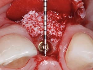
Connective Tissue and PRGF in Treatment of Periodontal Intrabony Defects
Description:
In order to reconstruct the lost periodontal tissues different kinds of graft materials have been used, including auto-grafts, allografts and xenografts and also Polypeptide growth factors in combination with guided tissue regeneration (GTR).In this presentation Dr Omid Moghaddas will present the concepts of using connective tissue and PRGF in treatment of intrabony defects and also review the literature on this topic.
Date Added:
1/29/2013
Author(s):
Omid Moghaddas, DMD, MSD
Dr Omid Moghaddas, DDS,MSD
Board Certified Periodontist
- Founder of Moghaddas Periodontics and dental implant center
- F...
[read more]
|

|
Online Videos / Surgery / Soft Tissue / Connective Tissue and PRGF in Treatment of Periodontal Intrabony Defects
Questions & Comments
|
omid moghaddas - (4/5/2013 12:20 AM)
Keith; by using PRGF bone fill in regeneration of periodontal defects was not significantly improved in conjunction with barriers or CTG,but using it with bone grafts without membranes was beneficial.i am agree with dr salama in positive role of prgf in improving soft tissue healing.tnx. Dr Moghaddas
|
Maurice Salama - (3/4/2013 7:06 AM)
Keith; I only utilize PRGF with ACDM CTG cases. But, it certainly has a positive effect on soft tissue wound healing. Dr. Salama
|
Keith Marshall - (3/3/2013 7:40 PM)
Am I correct in that if you are going to use a CT graft in any periodontal procedure the prgf will not improve the outcome in a significant way?
|
Related Videos |
|
|
Modern Crown Lengthening: Classification and Clinical Application
Crown lengthening procedures are indicated to provide adequate tooth structure in case of subgingival tooth fracture or caries, uneven gingival level, un-esthetic short crowns due to the tooth wear, inadequate axial height, altered passive eruption ,forced eruption of a single or multiple teeth and finally in case of gingival smile.
The treatment modality for esthetic crown lengthening procedure should be based on detail diagnosis in each case because of the multifactorial etiology and because of the type of therapy selected by the clinician, will have a direct implication for the esthetic result.
Presented By:: |
André P. Saadoun, DDS, MS |
Presentation Style: |
Video |
Community Rating: |
|
|
Watch Now>>
|
|
|
|
|
Esthetics Around Implants; Introducing the "Snake" Technique !
The “Snake technique” is a new concept in minimally invasive surgery.
It is based on old fundamental principles that offer reliable and predictable results. The “Snake technique” is a pediculated gingival graft, which unlike free grafts (CTG, FGG) offers several advantages and a lower surgical stress for our patients by having no need for an additional surgical site. As the small details make the biggest differences, the “Snake technique” may add value to both the surgical course of therapies selected as well as the relationship with our patients who demand minimally invasive treatments.
Presented By:: |
Cosmin Dima, DDS |
Presentation Style: |
Video |
Community Rating: |
|
|
Watch Now>>
|
|
|
|
|
A Defined Algorithm for Regenerative Success Part 2
These presentations will focus open the above concepts but prioritize the diagnostic phase and surgical common denominators required for a successful regenerative outcome. Flap design, space maintenance, bone and membrane selections and tension free closure provides the template for all the new age materials and technologies to be successful in clinical practice.
Presented By:: |
Maurice Salama, DMD |
Presentation Style: |
Video |
Community Rating: |
|
|
Watch Now>>
|
|
|
|
Related Courses |
|
|
Team Work: The Role of the Restorative Dentist in the Multidisciplinary Practice
In this presentation, Drs. Conti and Rossi share their experiences in treating challenging cases from a comprehensive multidisciplinary perspective. Emphasis will be placed as to when and how the restorative dentist enhances overall periodontal and esthetic therapy.
Presented By:: |
Roberto Rossi, DDS;Alessandro Conti, DDS |
Presentation Style: |
Online Self-Study Course |
CE Hours: |
1 CEU (Continuing Education Unit) |
|
Watch Now>>
|
|
|
|
|
Positive & Negative Factors in Stem Cells & Wound Healing: Solutions for Long Term Stability
The main factor for soft and hard tissue healing is the speed and quality of new vascularization.
This lecture will present all the positive and negative factors who control the angiogenesis, blood supply and bone metabolism:
-Biological factors as vit. D and cholesterol have to be checked before the surgery
-The management of the flaps with sutures is one the main factor for the bone and soft tissue maintenance..
-The soft brushing technique is a new technique which increases the flap without periosteal incision nor bleeding.
-Careful Implant placement without too much torque, specially in cortical bone or grafted bone at the re-entry..
-The A-PRF liquid and i-PRF are solutions to reduce the mobility of the granules (sticky bone) with an improvement of angiogenesis
The use of growth factors is a stimulation of angiogenesis.
The prevention of the negative factors will allow us to achieve the long term stability.
Presented By:: |
Joseph Choukroun, MD |
Presentation Style: |
Online Self-Study Course |
CE Hours: |
1 Continuing Education Unit (CEU) |
|
Watch Now>>
|
|
|
|
|
Microsurgical Soft Tissue Reconstruction for Teeth & Implants - Part 2 of 2
Esthetics plays a key role in a modern society and the average dental health of the population is improving every day. In such environment, the demands of dental treatments are different and dentists should approach their patients in a different way: they should be prepared to implement different and modern techniques around teeth and implants. Soft tissue grafting techniques provide the dentist with an armamentarium that makes possible to prevent and solve many problems and complications around teeth and implants, making more predictable the treatments results.
This presentation is focused in the importance of soft tissue grafting techniques around teeth and implants and how to do it in a predictable, modern and a minimally invasive way. We present many different techniques illustrated with clinical cases.
Presented By:: |
Ramon Gomez Meda, DDS |
Presentation Style: |
Online Self-Study Course |
CE Hours: |
1 CEU (Continuing Education Unit) |
|
Watch Now>>
|
|
|
|
Related Articles |
|
|
The Use of PerioDerm for Root Coverage and Correction of Insufficiently Attached Gingiva
The objective of this article is to demonstrate the benefits
of an acellular dermal matrix called PerioDerm™
Acellular Dermis in correcting recession and adding connective
tissue during periodontal-restorative comprehensive treatment.
1,2 It has long been established that connective tissue
is an integral factor in the protection of the underlying periodontal foundation. Connective tissue tends to be tenacious in makeup and have far less vascularization than mucosa. It prevents bacterial infiltration to the underlying supportive periodontal tissues, and in restorative applications, it adds stability to gingival areas during impression taking and cementation of restorations. Though the quality of tissue is critical, it is also important that the tissue is bound down to either tooth surface or bone to serve as a supportive mechanism for protection of the periodontium.
|
|
|
|
The Management of Immediate Implant Placement to Optimize Aesthetic Outcome in the Anterior Maxilla
Following tooth extraction the implant surgeon may select between various implant placement timing and loading protocols. Ideally these are to be determined prior to extraction, be it immediate, early, or late placement. Immediate implant placement even in the aesthetic zone is a literature supported treatment modality with success comparable to alternative placement protocols. Meticulous restorative treatment planning of a tooth destined
for extraction is essential. Selecting the appropriate implant and techniques may preserve and ensure natural aesthetics. Utilizing the patient’s own tooth crown can better provisionalize the implant with a ‘walk out as you walk in’
result.
Author(s): |
Howard Gluckman, BDS, MChD, PhD;Jonathan Du Toit |
|
View Article>>
|
|
|
|
|
Soft Tissue Cone-Beam Computed Tomography: A Novel Method for the Measurement of Gingival Tissue and the Dimensions of the Dentogingival Unit
The purpose of the
present study was to develop a soft tissue cone-beam computed tomography (ST-CBCT) to
improve soft tissue image quality and allow the determination of the dimensions and relationships
of the structures of the dentogingival unit. Two separate CBCT scans were obtained from
three patients with different periodontal biotypes. The first was a scan following standard
methods; however, for the ST-CBCT the patients wore a plastic lip retractor and retracted their
tongues toward the floor of their mouths.
Author(s): |
ALESSANDRO LOURENÇO JANUÁRIO, DDS, MS, PhD; MAURÍCIO BARRIVIERA, DDS, MS; WAGNER RODRIGUES DUARTE, DDS, PhD |
|
View Article>>
|
|
|
|
|