Video Details
|
Dental Photography: From Theory to Practice
Description:
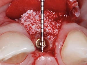
Dental photography should be your daily tool. If not, don’t wait and take your steps to learn it and master it. In this lecture, you will see how to take step by step full dental portfolio and which instruments are helpful in surgical photo documentation. In the end, you will get an update in settings, equipment, and accessories used to take excellent dental photo documentation.
Date Added:
7/8/2020
Author(s):
Kris Chmielewski, DDS, MSc
Krzysztof "Kris" Chmielewski, DDS, MSc
- 1993 – Graduated Medical Academy in Gdansk
- 1993 -1994 Assistant in Prosthetic Department ...
[read more]
|

|
Online Videos / General / Other Specialties / Dental Photography: From Theory to Practice
Questions & Comments
|
Raymond La Vigne - (7/8/2020 10:09 PM)
very excellent photography my suggestion divide duties only either assistant or Doctor wears gloves and contacts patient and retractors other ungloved person handles extraoral camera much lower or zero chance of cross contamination for patient or camera
|
Vijay Kumar - (5/20/2018 1:42 AM)
Great lecture with very practical explanations.
dr kris .
i want to know which co. is selling mirrors and contrasters you have used in this presentation
|
Raymond La Vigne - (4/11/2018 7:59 PM)
great photography but poor use of gloves and infection control I saw significant cross-contamination Would be better to have assistant be gloved and touch everything but camera and doctor have clean ungloved hands and only touch camera
|
Tomas Vanek - (4/11/2018 6:14 PM)
Thanks a lot Kris! It is wonderful photo-tour. I am sure I will be the full member of your web site "photo dental masters" - it looks great! It is fantastic to see somebody from the middle Europe as I am.... sincerely, Tomas, Czech Republic
Maurice - hairdryer a few seconds before - it is well done for couple of minutes
|
Maurice Salama - (4/11/2018 9:17 AM)
Well done. How do you prevent fog in the mirrors intra-orally?
|
Related Videos |
|
|
Non-Surgical Management of Progressive Condylar Resorption & Degenerative Joint Disease
This presentation does an overview of PCR/DJD in young female patients that is secondary to the use of ethinyl estradiol birth control pills.
An overview of the pathophysiology and medical management as a component in the treatment of PCR/DJD is presented.
There are several case reviews demonstrating diagnostic and non-surgical treatment protocol for non-surgical management of PCR/DJD.
Closing recommendations for implementing early recognition and patient education of this condition are also provided.
Presented By:: |
Brian C. Mills, DMD |
Presentation Style: |
Video |
Community Rating: |
|
|
Watch Now>>
|
|
|
|
|
|
Analogue vs. Digital Dentistry
In the progressive world of modern digital technologies, it is more apparent than ever that there is always an analogue component to the restorative clinical process. This presentation will discuss the diagnostic aspects of restorative based digital dentistry as well as the accuracy advantages to the final outcomes. Digital Smile planning, smile mockups and their critical role in minimally invasive tooth preparation will be highlighted. Lab communication with these digital templates relay all the necessary information and provide the lab technician with a more manageable workflow. Accuracy, better outcomes and less invasive dentistry are the winners in this evolution.
Presented By:: |
Nazariy Mykhaylyuk, DMD |
Presentation Style: |
Video |
Community Rating: |
|
|
Watch Now>>
|
|
|
|
Related Courses |
|
|
Dental Photography: From Theory to Practice
Dental photography should be your daily tool. If not, don’t wait and take your steps to learn it and master it. In this lecture, you will see how to take step by step full dental portfolio and which instruments are helpful in surgical photo documentation. In the end, you will get an update in settings, equipment, and accessories used to take excellent dental photo documentation.
Presented By:: |
Kris Chmielewski, DDS, MSc |
Presentation Style: |
Online Self-Study Course |
CE Hours: |
1 CEU (Continuing Education Unit) |
|
Watch Now>>
|
|
|
|
|
|
From 2D to 3D: The Benefits of 3D-X-ray in the Different Fields of Dentistry
CBCT(Cone Beam Computed Tomography) has developed in the last years from a highly specialized used subject used mainly by specialized surgeons and implantologists to a wide field of indications, used mostly by general practitioners, periodontists, endodontists, general implantologists and orthodontists. The picture quality has increased drastically so the 3-D X-ray techniques are able to show aspects that 2-D X-ray-techniques are unable to present. 3-D X-ray systems are linked today to other diagnostic and electro-mechanical systems as there are automaticised software measure and diagnostic tools, intra and extra-oral scanning systems and CAD-CAM-milling-systems. That increases the diagnostic and treatment possibilities this systems offers to the users.
Presented By:: |
Robert Kalla, Dr. med., dent. |
Presentation Style: |
Online Self-Study Course |
CE Hours: |
1 CEU (Continuing Education Unit) |
|
Watch Now>>
|
|
|
|
|
The Digital Dental System: Intra-Oral Scanning and Design
This webinar is designed for the clinician who desires to learn and use digital acquisition scanners. This is the future of any dental practice and it all starts by sitting at the computer and designing your crowns and veneers. This course will help the beginner and advanced digital clinician. The aspects of preparation design, aesthetics and occlusion will be demonstrated. If you own a digital scanner or thought of purchasing one, this is the course that will show you the real utilization of this million dollar technology and increase your bottom line for your practice.
Presented By:: |
Dean C. Vafiadis, DDS |
Presentation Style: |
Online Self-Study Course |
CE Hours: |
1 CEU (Continuing Education Unit) |
|
Watch Now>>
|
|
|
|
Related Articles |
|
|
|
Laboratory Technology
Prosthodontic treatment ofthe edentulous cleft palate patient presents the dentist with numerous challenges for achieving a satisfactory result for the patient. A technique is described for prosthodontic rehabilitation of a completely edentulous cleft palate patient using bone augmentation procedures, root-form dental implants, and a removable maxillofacial prosthesis. Dental implants may improve prosthesis retention, stability, and occlusal function when used in carefully selected cases.
Author(s): |
Lee H. Silverstein, DDS, MS, FACD, FICD;Michael D. Lefkove, DDS, Bill Matheny, CDT |
|
View Article>>
|
|
|
|
|
|