Video Details
|
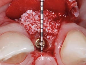
Digital Dynamic & Static Solutions in Partial Extraction Therapy
Description:
Partial Extraction Therapy has changed the methods of implant planning and execution. Preserving both hard and soft tissue has propelled these concepts to innovative. Until recently the use of dynamic and static guidance has been delayed. The objective of this presentation is to understand the need for digital guidance and explores avenues to accomplish a digitally based workflow and execution fr the subsequent procedures.
Date Added:
10/21/2020
Author(s):
Isaac D Tawil, DDS
Dr. Isaac Tawil received his Doctor of Dental Surgery Degree from New York University College of Dental Surgery and has an Masters degree in Biology.
Dr. Ta...
[read more]
|

|
Online Videos / Surgery / Other Surgical / Digital Dynamic & Static Solutions in Partial Extraction Therapy
Questions & Comments
|
Isaac Tawil - (11/1/2020 4:49 PM)
Thank you guys for the comments 🙏
|
Isaac Tawil - (11/1/2020 4:49 PM)
Thank you Snjezana. We are working on getting TBS in Europe. I’m hoping to have by the end of the year. Currently Cguide is nit in R2 library but choosing standard r2 5mm guide will for medium G stops.
|
richard martin - (10/23/2020 10:05 PM)
ITAL,
Always pushing forward - as you know - big fan of the stackable - and thank you for sharing and dedication to guided as snazzy mentioned - keep rolling- Snazzy # shape library being modified
Cheers
|
snjezana pohl - (10/21/2020 12:50 PM)
Dear Isaac, what to say: I feel so old-fashioned watching your accomplishments! Your contribution to the guided socket shield preparation is tremendous!
Are TBS instruments available in Europe?
Are c-shape Densah guides in R2 Gate library? I am asking because despite all efforts from Versah site, 3-shape still does not offer the possibility to utilize c-shape guides.
And, Isaac, is there a manual for stacks production for dummies?
Keep leading and innovating!
|
Charles Schwimer - (10/20/2020 8:25 AM)
Just Wow! Masterful blending of the Clinical and Technological worlds. The future is coming fast thanx to your efforts. Congratulations my friend!
|
Related Videos |
|
|
Implant Removal Made Easy - When Peri-implantitis Treament is Not Possible
In this video and surgical presentation, Dr. Eduardo Anitua demonstrates a step-by-step safe, efficient and predictable surgical/mechanical technique for the removal of implants that are severely compromised by peri-implantitis. In addition, Dr. Anitua will discuss how to manage the regeneration of the site following implant removal through the therapeutic utilization of plasma rich in growth factors, i.e. Endoret.
Presented By:: |
Eduardo Anitua, MD, DDS, PhD |
Presentation Style: |
Video |
Community Rating: |
|
|
Watch Now>>
|
|
|
|
|
Venipuncture & Blood Drawing: Phlebotomy Step-by-Step
The use of platelets concentrates is becoming unavoidable in our surgeries.
To take full advantage of these techniques, the surgeon has to be familiar with the blood collection technique and capable to draw blood at any time. This presentation will show you how to manage simply the venipuncture according to a step by step protocol, with special attention for tricky situations & difficult patients.
Presented By:: |
Elisa Choukroun, DDS |
Presentation Style: |
Video |
Community Rating: |
|
|
Watch Now>>
|
|
|
|
|
Lasers in Contemporary Implant and Restorative Dentistry: A Microscopic Perspective
In the lecture, we will discuss the role of lasers after the implant has been placed. We will demonstrate how it can be used to improve healing. It can be used during uncovery. It can be used in periimplant tissue removal, and even in the exciting world of periimplantitis. Clinical cases will be presented showing hard-tissue lasers being used for decortication as an alternative to traditional means. We will focus on why an electrosurge may be damaging to your implants, and may cause you more problems than benefits, and why a diode-laser may be your treatment of choice.
Presented By:: |
Glenn A. van As, BSc, DMD |
Presentation Style: |
Video |
Community Rating: |
|
|
Watch Now>>
|
|
|
|
Related Courses |
|
|
The Surgical-Restorative Digital Work Flow In Comprehensive Therapy
Traditional methods of treating our patients are being been replaced by digital processes, impacting our daily life. The intake of various technologies such as CBCT imaging, scanning, digital restorative design and CAD/CAM production have contributed to this movement. In the same time, restorative materials have also been developed and studied that have improved strength and esthetic qualities. All these capabilities create opportunities for better restorative outcome.
This presentation will highlight a practice-based model centered on digitally enhanced treatment for efficient and predictable esthetic restorative outcomes.
Presented By:: |
Karim Dada, DDS, MS;Leon Pariente, DDS |
Presentation Style: |
Online Self-Study Course |
CE Hours: |
1 CEU (Continuing Education Unit) |
|
Watch Now>>
|
|
|
|
|
Next Generation Biomaterials for Bone and Periodontal Regeneration - Part 1 of 2
New and innovative biomaterials are being discovered or created in laboratories at an unprecedented rate, but many entirely unknown to practicing clinicians. This course addresses this gap in knowledge by summarizing some of the groundbreaking research performed to date on this topic and provides case examples of these biomaterials at work. The course begins with a review of the biologic background and applications of bone grafting materials utilized in dentistry. Thereafter, new and exciting updates on Platelet Rich Fibrin, Emdogain/Osteogain, Tetranite and Vitamin D deficiency are discussed with respect to their ability to promote either bone or periodontal regeneration.
Presented By:: |
Richard Miron, DDS, BMSC, MSc, PhD |
Presentation Style: |
Online Self-Study Course |
CE Hours: |
1 CEU (Continuing Education Unit) |
|
Watch Now>>
|
|
|
|
|
Piezosurgery on Impacted Teeth
In the latest years, Oral surgery has strongly suffered of the technological innovations introduced in this field. In particular, the use of ultrasound applied to surgery has changed some of the most frequent clinical procedures, such as the extraction of third parties molars included, thus spreading an innovative concept in all dentistry: piezoelectric surgery or piezosurgery. So taking care of about 10 years of simple and complex extractive surgery I had the need to elaborate a lecture in which they are described in a simple and linear way the surgical techniques for the avulsion of the dental elements included, to provide a repeatable and predictable approach and method in order to reduce their invasiveness and traumaticity procedures
Presented By:: |
Prof. Dott. Angelo Cardarelli |
Presentation Style: |
Online Self-Study Course |
CE Hours: |
1 CEU (Continuing Education Unit) |
|
Watch Now>>
|
|
|
|
Related Articles |
|
|
A Technique for Surgical Mandibular Exostosis Removal
Exostosis, a slow-growing, benign bony outgrowth, is a common clinical finding and not usually an issue with patients. However, when removable prosthetics must sit either adjacent to or over these areas, pressure, food abrasion, ulceration, or limited tongue space can occur. This article describes a surgical technique for exdsion of exostosis through the presentation of a case. An 86-year-old woman had soft-tissue irritation caused by abrasion from food in the buccal posterior right quadrant. The…
Author(s): |
Lee H. Silverstein, DDS, MS, FACD, FICD;Gregori M. Kutzman, DDS |
|
View Article>>
|
|
|
|
|
Applied Techniques for Predictable Suture Placement Part 1
Surgical suture positioning is crucial to ensure adequate healing and can be accomplished using a variety of suturing methods. Sutures should generally be placed distal to the last tooth and within each interproximal space and should always be inserted through the more mobile flap first The flaps should not be blanched during the tying procedure, and closure should not be positioned closer than 2 mm to 3 mm from the edge of the flap, in order to prevent tearing during the inevitable swelling that…
|
|
|
|
The Socket-Shield Technique: First Histological, Clinical, and Volumetrical Observations after Separation of the Buccal Tooth Segment – A Pilot Study
The “socket-shield technique” has shown its potential in preserving buccal tissues. However, front teeth often
have to be extracted due to vertical fractures in buccolingual direction. It has not yet been investigated if the socket-shield
technique can only be used with intact roots or also works with a modified shield design referring to vertical fracture lines.
The aim of this study was to assess histologically, clinically, and volumetrically the effect of separating the
remaining buccal root segment in two pieces before immediate implant placement.
Author(s): |
Markus B. Hürzeler, DMD, PhD;Daniel Bäumer, DDS; Otto Zuhr, DDS; Stephan Rebele, DDS; David Schneider, DDS, PhD; Peter Schupbach, PhD |
|
View Article>>
|
|
|
|
|