Video Details
|
Effective Techniques for Placement of Narrow Diameter Implants - Part 2 of 3
Description:
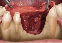
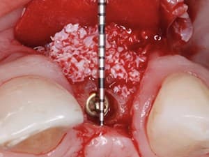
This presentation utilizes results from a prospective clinical trial as well as clinical experience to outline indications for effective and efficient utilization of narrow diameter implants in regions of limited space. Along with a simplified approach to placement, soft tissue management and provisionalization will be discussed in depth.
Date Added:
7/1/2011
Author(s):
Michael Reddy, DMD, DMSc
Dr. Reddy is Professor and Chairman of the Department of Periodontology at the University of Alabama at Birmingham School of Dentistry. Dr. Reddy is a 1981 graduate of...
[read more]
|

|
Online Videos / Surgery / Implant / Effective Techniques for Placement of Narrow Diameter Implants - Part 2 of 3
Questions & Comments
|
Ashwath Gowda - (7/7/2012 3:11 PM)
You have preared the post for the crown on that right mxillary lateral RIGHT AFTER placing the implant.
First of all... no matter, how much of care we exert NOT to disturb the initial "INTEGRITY" (torqued)between bone and the tiny narrow implant...the implant is most likely to "spin" during this "un-timely" procedure!!!!
Second...How are those flying metallic grind particles prevented or controlled from entering and ebeddig into the raw soft tissue at the surgical site???
|
Maria Jose Iturburu - (7/2/2011 8:29 AM)
very good tecnique to an unresolving problem
|
Related Videos |
|
|
|
|
The Central-Lateral Replacement Dilemma in Esthetic Implant Dentistry: Clinical Alternatives & Case Management
Clinicians today have access to an astounding array of new technology, tools and materials to utilize in their progressive and esthetically oriented practices. However, none of them can ensure success if the treatment design is inadequate. This presentation puts an emphasis on identifying the risk factors to successful esthetic outcomes specific to multiple adjacent tooth replacement within the Esthetic Zone. Specifically, the challenges to replacement of adjacent Maxillary Lateral and Central Incisors will be highlighted as well as developing appropriate solutions to overcoming them.
Presented By:: |
Maurice Salama, DMD |
Presentation Style: |
Video |
Community Rating: |
|
|
Watch Now>>
|
|
|
|
|
Zirconia Implants in the Age of 3D Imaging: An Alternative Therapy for Aesthetics and Function
Computer Guiding dental implants provides perfect positioning for mechanics and cosmetic results. The advantages provided by the implant selection utilizing interactive software libraries allows the implantologist to choose the best diameter, length and implant design for the specific surgical site. Zirconia implants can be computer guided and planned with interactive software and will provide enhanced aesthetic results as well as top gingival health due to the multiple advantages of zirconia.
Presented By:: |
Alvaro Ordoñez, DDS |
Presentation Style: |
Video |
Community Rating: |
|
|
Watch Now>>
|
|
|
|
|
Related Courses |
|
|
Total Rehabilitation of the Edentulous Patient with the All-on-4® treatment concept
A fixed implant supported prosthesis is the ideal treatment option for the edentulous patient and those facing the imminent loss of their remaining dentition. Through innovation, along with continuous clinical and scientific research, it has become possible to treat virtually every edentulous case with a highly functional and esthetic fixed solution. The oral rehabilitation of these patients has two stages: surgical and prosthetic.
Presented By:: |
Paulo Malo, DDS, PhD |
Presentation Style: |
Online Self-Study Course |
CE Hours: |
1 CEU (Continuing Education Unit) |
|
Watch Now>>
|
|
|
|
|
Esthetic Zone Reconstruction: Synergy of Hard and Soft Tissue Grafting
The loss of alveolar ridge contour in the esthetic zone compromises both esthetics and function. This clinical presentation will focus on the application of both hard and soft tissue grafting in the esthetic zone implementing state of the art interactive CT diagnosis and treatment planning for optimal esthetic implant reconstruction.
Emphasis will be on indications, graft and harvest site assessment, timing, and use of bioactive modifiers including BMP, PRGF and PDGF. The surgical protocol utilizing allogeneic and autogenous grafts in conjunction with connective tissue grafts, acellular dermis matrix, and related soft tissue procedures to avoid functional and esthetic pitfalls will be featured. Pre and post-grafting prosthetic workup, interactive CT graft assessment and perio prosthetic driven implant planning, stent driven implant placement, and computer milled abutment fabrication including guided surgery will be included.
Presented By:: |
|
Presentation Style: |
Online Self-Study Course |
CE Hours: |
1 CEU (Continuing Education Unit) |
|
Watch Now>>
|
|
|
|
|
Surgical Management of Posterior Maxilla
Replacement of missing teeth with dental Implants in the posterior maxilla presents unique challenges due to maxillary sinus, less quality of bone, and increased occlusal load. The surgical objectives to meet functional and aesthetic goals include enhancement of both quantity and quality of bone as well as ideal placement of dental implants for restorations that mimic nature. In this presentation, Dr. H. Ryan Kazemi discusses current surgical techniques in implant site development in the posterior maxilla including extraction site grafting, sinus lift, bi-directional bone graft, and osseodensification for bone management.
Presented By:: |
H. Ryan Kazemi, DMD |
Presentation Style: |
Online Self-Study Course |
CE Hours: |
1 Continuing Education Unit (CEU) |
|
Watch Now>>
|
|
|
|
Related Articles |
|
|
Diameter Selection of Implants Placed in Extraction Sockets: A New Approach
Implant placement in extraction sockets is considered a daunting challenge
due to offering risks of bone remodeling and consequent gingival alterations. Objective: This
study aims at proposing a protocol for selecting the diameter of upper anterior osseointegrated
implants placed in extraction sockets. This protocol was based on the bucco-palatal dimension of
the socket and allows a 3-mm gap to form between the implant and the outer surface of the buccal
bone wall. Such gap must be filled with biomaterial or autograft so as to increase the predictability
of long-term results.
Author(s): |
José Carlos Martins da Rosa, DDS, MS;Ariádene Cristina Pértile de Oliveira Rosa; Carlos Eduardo Fra ncischone; Bruno Salles Sotto-Maior |
|
View Article>>
|
|
|
|
|
Success Rate, Healing Time, and Implant Stability of Photofunctionalized Dental Implants
This is the first study to report the clinical outcomes of photofunctionalized dental implants. This retrospective study analyzed 95 consecutive patients who received 222 untreated implants and 70 patients who received 168 photofunctionalized implants over a follow-up period of 2.5 years. Photofunctionalization was performed by treating implants with UV light for 15 minutes using a photo device
immediately before placement. Within the
limits of this retrospective study, despite the more frequent use of shorter and smaller-diameter implants, the
use of photofunctionalization allowed for a faster loading protocol without compromising the success rate.
Author(s): |
Akiyoshi Funato, D.D.S.;Masahiro Yamada; Takahiro Ogawa |
|
View Article>>
|
|
|
|
|
|