Video Details
|
複雑な抜歯部位におけるGBR
Description:
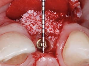
In this surgical technique video, Dr. Michael Sonick shares a routine clinical challenge and demonstrates the extraction of a fractured premolar with simultaneous guided bone regeneration. His very detailed and concise demonstration focuses on the preparation of the extraction site as well as the utilization of growth factors and membranes.
Date Added:
9/24/2010
Author(s):
Michael Sonick, DMD
Dr. Michael Sonick is a full time practicing periodontist and implant surgeon in Fairfield, Connecticut. He is also an active teacher, clinical researcher and author. ...
[read more]
|

|
Online Videos / Surgery / Bone Grafting / Guided Bone Regeneration of a Compromised Extraction Site
Questions & Comments
|
Joon Kim554 - (10/7/2017 9:33 AM)
Do you use surgical handpiece with the brassler bur 7408028? Is this a carbide bur that people often use for their composite?
|
Joon Kim554 - (10/7/2017 9:31 AM)
I wouldn't cover the top part of the socket with the membrane you used on covering the buccal. All resorbable membranes (even the long lasting ones), resorb too early when exposed to saliva enzyme. These membranes are supposed to be used for primary closure but we need secondary closure to achieve keratinized tissue. Ideal way to do it is do exactly what you did but also cover the top part with PTFE non resorbable membrane.
|
alireza torabi - (8/10/2013 9:06 PM)
Good job!
|
Mike Sonick - (4/20/2013 11:19 AM)
George. Thanks for your question. Please see my answer below to Benjamin. I agree with the publication by Artzi. That is why I use cancellous allograft with a growth factor. I obtain 50% vital bone in 6 months.
|
Mike Sonick - (4/20/2013 11:17 AM)
Benjamin
There is not way to know if there is vital bone without a biopsy. In my experience the use of BioOss and Biogide results in 50% vital bone in approximately 6-9 months. The turnover is slow. I usually use Puros or some other bone allograft with Gem 21 and a collagen membrane. I have been able to obtain 50% vital bone in 4 months with this technique. For more information you may want to consult my text Implant Site Development published by Wiley Blackwell
|
Benjamin Saidel - (2/21/2013 8:36 PM)
Dr Sonick I am a quite new with these bone grafting techniques so your assistance will be greatly appreciated
I grafted a socket following the extraction of a upper left first molar last year which had fractured post endo. (There was periapical pathology extending into the furcation with an associated buccal swelling.)
Following extraction of the tooth it became obvious that the buccal plate had been lost so I raised a flap and grafted the socket using only Bio-OSS and a Bio-guide membrane. The socket healed uneventfully ( some granules had leaked out from under the membrane at 1 week review but did not seem to adversely affect healing) This was 6- 8 months ago. I have reviewed her a couple of times during this period and clinically the tissues have healed well. I have also taken peri apical X-rays which show the graft material
The patient is now ready to have the implant placed. I have taken a CBCT and in the area of the socket is a dense radiopacity which must be the graft material. My question to you is the following- Has bone formed in this area or is it only graft material?
I am now concerned that when I raise a flap to place the implant all I will find is graft material with little or no new bone,which may even come away in the flap and I will be left with an open socket or partially healed socket
Kind regards
Benjamin Saidel
London
|
George Kotsakis - (4/24/2011 11:19 AM)
Dr Sonick, you have been fastiduous in explaining us every step as always, thank you very much. I have only one question, you seem to have used some bovine graft as well. I have in my mind the publication by Artzi et al on bovine xenografts, where after 6 months the had minimal mature bone, mostly CT on the coronal aspect of the socket and almost 35% of xenograft all over the socket. Howcome u manage to get back in, as early as 5 months and get dense bone???
|
Mike Sonick - (12/12/2010 6:12 PM)
Jorge The membrane was not exposed. It was placed underneath the tissue and a collagen plug placed coronally. altho the collagen plug is left exposed this does not seem to be a problem for it is completely epithelialized within two weeks. I used to use CTG for this procedure but have found them to take more time with no improvement in result.
Immediate implants without a flap with this technique would be a probelm for the buccal plate would not be regenerated. It is all about eh buccal plate. If no buccal plate exists you must flap and regenerate independent of whether you are placing an implant or not.
|
Jorge Gutierrez - (11/29/2010 9:42 AM)
Dr Michael I would like to know in your video the membrane was exposed to the mouth that does not compromise the final result of their work, would not be better to cover with tissue graft? What do you think of making an immediate implants with the technique without flap?
|
|
Related Videos |
|
|
Vertical Bone Augmentation: Current Concepts and Techniques
This lecture will review several approaches used for vertical bone augmentation and discuss the benefits and limitations of each. There will be special emphasis on the titanium mesh and inter-positional osteotomy techniques.
Presented By:: |
David Dara Yarmand, DDS, MD |
Presentation Style: |
Video |
Community Rating: |
|
|
Watch Now>>
|
|
|
|
|
|
Restoration of Optimal Esthetics in Complex Clinical Situations
Patients often neglect or delay dental care for an extended time because of their negative experiences with and fear of dental treatments. This neglect may result in the advance of periodontitis with severe bone loss, a substantial number of missing teeth, and problems with the remaining teeth. Preexisting malocclusion can aggravate this condition. This case report
demonstrates an interdisciplinary strategic approach, in combination with the use of biologically grounded, clinically proven, and viable techniques and technologies to restore optimal esthetics and function in such complex cases. The authors emphasize the importance of carefully planning the timing and sequence of performing periodontal, orthodontic, and implant therapies. The advanced techniques and technologies successfully implemented in this case report include the growth factor and titanium mesh (Ti mesh)-assisted bone augmentation; ovate pontic-mediated, noninvasive soft tissue contouring; and photofunctionalization
Presented By:: |
Tomohiro Ishikawa, DDS |
Presentation Style: |
Video |
Community Rating: |
|
|
Watch Now>>
|
|
|
|
Related Courses |
|
|
A GRAFT-LESS Approach to Implant Dentistry
Vertical bone augmentation techniques are more invasive and success rates often vary according to the surgical expertise of the clinician. There has been a trend over time towards minimally invasive treatment options in implant dentistry. This approach attempts to avoid more complicated procedures involving bone augmentation for implant placement. This presentation discusses a “graft less” treatment philosophy that emphasizes the use of less demanding augmentation techniques for the purpose of placing short implants in atrophic posterior sites.
Presented By:: |
Craig M Misch, DDS, MDS |
Presentation Style: |
Online Self-Study Course |
CE Hours: |
1 CEU (Continuing Education Unit) |
|
Watch Now>>
|
|
|
|
|
Titanium Mesh and rhBMP-2 in Ridge Augmentation: Options and Limitations
Titanium mesh has been used in reconstructive surgery and ridge augmentation for several years. The traditional technique involves using cancellous autogenous bone under the mesh scaffold. More recently growth factors and bone substitutes have been utilized as autograft replacements. There are several advantages to using titanium mesh for space maintenance including strength, ease of use, low cost and biocompatibility. However, the difficulty in removing the mesh has led to a search for other space maintenance scaffolds including tenting screws, dense preshaped collagen and porous high-density polyethylene. This webinar will discuss the use of rhBMP-2 for managing horizontal and vertical bone deficiencies with various scaffolds.
Presented By:: |
Craig M Misch, DDS, MDS |
Presentation Style: |
Online Self-Study Course |
CE Hours: |
1 CEU (Continuing Education Unit) |
|
Watch Now>>
|
|
|
|
|
Esthetic Zone Reconstruction: Synergy of Hard and Soft Tissue Grafting
The loss of alveolar ridge contour in the esthetic zone compromises both esthetics and function. This clinical presentation will focus on the application of both hard and soft tissue grafting in the esthetic zone implementing state of the art interactive CT diagnosis and treatment planning for optimal esthetic implant reconstruction.
Emphasis will be on indications, graft and harvest site assessment, timing, and use of bioactive modifiers including BMP, PRGF and PDGF. The surgical protocol utilizing allogeneic and autogenous grafts in conjunction with connective tissue grafts, acellular dermis matrix, and related soft tissue procedures to avoid functional and esthetic pitfalls will be featured. Pre and post-grafting prosthetic workup, interactive CT graft assessment and perio prosthetic driven implant planning, stent driven implant placement, and computer milled abutment fabrication including guided surgery will be included.
Presented By:: |
|
Presentation Style: |
Online Self-Study Course |
CE Hours: |
1 CEU (Continuing Education Unit) |
|
Watch Now>>
|
|
|
|
|