Video Details
|
Immediate Dentoalveolar Restoration
Description:
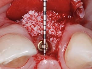
The IDR (Immediate Dentoalveolar Restoration) technique has been used for over 6 years. With it, we can reconstruct socket tissue losses in the same surgical session of dental extraction, implant placement and provisional crown installation, minimizing the treatment time and morbidity of the procedures.
In this presentation we discuss the hard and soft tissues stability when the IDR technique is applied in different types of periodontal biotypes and in sites with different defect levels.
The clinical, radiographic and tomographic follow-up of some cases is shown, allowing us to evaluate the maintenance of gingival architecture and peri-implant bone stability around the implants.
Date Added:
10/16/2013
Author(s):
José Carlos Martins da Rosa, DDS, MS
Dr. José Carlos Martins da Rosa received his DDS degree in 1988 from the Federal University of Santa Maria/RS - Brazil. In 1992 he got a post graduate certificate in Pe...
[read more]
|

|
Online Videos / Surgery / Bone Grafting / Immediate Dentoalveolar Restoration
Questions & Comments
|
ali sojoodi - (6/27/2018 8:47 AM)
https://drsojodi.com/
|
ali sojoodi - (6/23/2018 1:14 AM)
[url=https://drsojodi.com/all-about-implant]ایمپلنت دندان[/url]
[url=https://drsojodi.com/Laminated-teeth]لمینت دندان[/url]
[url=https://drsojodi.com/dental-implant-insertion]ایمپلنت[/url]
[url=https://drsojodi.com/Correction-of-smile-design]اطلاح طرح لبخند[/url]
[url=https://drsojodi.com/pull-tooth]کشیدن دندان[/url]
[url=https://drsojodi.com/pull-tooth] دندان عقل[/url]
[url=https://drsojodi.com/Correction-of-smile-design]طراحی لبخند[/url]
[url=https://drsojodi.com/veneer]کامپوزیت ونییر[/url]
[url=https://drsojodi.com/Scaling-teeth]جرم گیری[/url]
[url=https://drsojodi.com/Gum-Surgery]جراحی لثه[/url]
[url=https://drsojodi.com/Laugh-beauty]زیبایی خنده[/url]
[url=https://drsojodi.com/Laugh-beauty]زیبایی لبخند[/url]
[url=https://drsojodi.com/clinic]کلینیک دندانپزشکی[/url]
[url=https://drsojodi.com/The-best-dentist-in-west-area-of-Tehran]بهترین دندانپزشک [/url]
|
ali sojoodi - (6/23/2018 1:14 AM)
<a href="https://drsojodi.com/all-about-implant">ایمپلنت دندان </a>
<a href="https://drsojodi.com/Laminated-teeth">لمینت دندان </a>
<a href="https://drsojodi.com/dental-implant-insertion">ایمپلنت </a>
<a href="https://drsojodi.com/Correction-of-smile-design">اصلاح طرح لبخند </a>
<a href="https://drsojodi.com/pull-tooth">کشیدن دندان عقل </a>
<a href="https://drsojodi.com/pull-tooth">کشیدن دندان </a>
<a href="https://drsojodi.com/Correction-of-smile-design">طراحی لبخند </a>
<a href="https://drsojodi.com/veneer">کامپوزیت ونیر </a>
<a href="https://drsojodi.com/Scaling-teeth">جرم گیری </a>
<a href="https://drsojodi.com/Gum-Surgery">جراحی لثه </a>
<a href="https://drsojodi.com/Laugh-beauty">زیبایی خنده </a>
<a href="https://drsojodi.com/Laugh-beauty">زیبایی لبخند </a>
|
benjamin yan - (1/25/2014 4:19 AM)
Dr.Jose,impressive presentation!Thank you.
|
Maurice Salama - (10/22/2013 7:16 AM)
Ganesh; I believe that the graft is stabilized enough by placement of the implant and compression from outer flap. Also, the highly vascular nature of this bone graft being mostly cancellous bone should help. Regards Dr. S
|
GANESH NAYAK - (10/21/2013 2:03 PM)
can the graft survive without stabilization
|
Márcia Souza951 - (10/20/2013 10:18 AM)
Congratulation.Amazing results.
|
Jose Carlos Rosa - (4/28/2013 6:27 PM)
Paul Boulos - First of all for this type of bone reconstruction the maxillary tuberosity is the preferred area for graft because their biological properties.
In cases with the presence of wisdom teeth and low bone availability, we can harvest bone of the lateral tuberosity. For this, we must have a cone beam computed tomography to know exactly and best area to be removed.
|
|
Related Videos |
|
|
|
|
Bone Augmentation & Vascular Enhancement Concepts: It’s Time to Make it Simple!
The purpose of this presentation is to make the participant think biology first and not mechanic first. A biological thinking will dramatically simplify vertical and/or horizontal bone augmentation procedures. We will also be introducing a new approach for the simplification of soft tissue handling: the Soft Brushing Technique, that will allow the coronal advancement of both buccal and lingual flaps, but also the palatal flap without the need for periosteal releasing incisions.
Presented By:: |
Jérôme Surmenian, DDS |
Presentation Style: |
Video |
Community Rating: |
|
|
Watch Now>>
|
|
|
|
Related Courses |
|
|
The Modern Age of Regeneration: Tenting, Fixating and Space Creation
Regeneration requires a common biological thread for predictable results. Space Maintenance, Graft Stability, Bone Biologics, and Wound Management. This presentation will describe the importance of each critical step in regeneration and introduce a new technical system to assist the surgeon in managing and stabilizing the space required for optimal bone regeneration. Flap design, release, biologics selection and enhancement as well as bone stabilization, fixation, and secure membrane adaptation will be further highlighted as to it's critical value in the final results.
Presented By:: |
Homa Zadeh, DDS, PhD |
Presentation Style: |
Online Self-Study Course |
CE Hours: |
1 CEU (Continuing Education Unit) |
|
Watch Now>>
|
|
|
|
|
Gaining Horizontal and Vertical Bone with Mineralized Bone Particulate, rh-BMP2 and Resorbable Ultrasonic-Applied Rigid Barriers
The use of titanium mesh has been well described, but so has the complications of removal and exposure. Bioresorbable barriers are now available which can be molded into any desired three-dimensional form, creating a rigid and resorbable construct that is secured to to the bone using ultrasonic energy. This unique solution has been successfully used to create the proper environment for both large lateral and vertical ridge augmentations for situations previously only treated with block grafting or distraction. Dr. Cummings will share his experience as well as the key soft tissue techniques that are critical for the success of any large augmentation.
Presented By:: |
Lewis C. Cummings, DDS, MS |
Presentation Style: |
Online Self-Study Course |
CE Hours: |
1 CEU (Continuing Education Unit) |
|
Watch Now>>
|
|
|
|
|
Bone Augmentation & Vascular Enhancement Concepts: It’s Time to Make it Simple!
The purpose of this presentation is to make the participant think biology first and not mechanic first. A biological thinking will dramatically simplify vertical and/or horizontal bone augmentation procedures. We will also be introducing a new approach for the simplification of soft tissue handling: the Soft Brushing Technique, that will allow the coronal advancement of both buccal and lingual flaps, but also the palatal flap without the need for periosteal releasing incisions.
Presented By:: |
Jérôme Surmenian, DDS |
Presentation Style: |
Online Self-Study Course |
CE Hours: |
1 Continuing Education Unit (CEU) |
|
Watch Now>>
|
|
|
|
Related Articles |
|
|
A Novel Strategy for Bone Integration and Regeneration: Case Studies
Ultraviolet light treatment of dental implants immediately prior to placement, or photofunctionalization, is a novel clinical tool with the potential to improve implant therapy. Photofunctionalization improves the surface properties of titanium surfaces by removing hydrocarbons, regenerating hydrophilicity, and optimizing electrostatic properties. We photofunctionalizated dental implants and titanium mesh (Ti mesh) in two complex clinical cases requiring simultaneous guided bone regeneration, sinus elevation, immediate implant placement into the extraction socket, and esthetic consideration
Author(s): |
Akiyoshi Funato, D.D.S.;Ryohei Tonotsuka, DDS; Hitochi Murabe, DDS; Makoto Hirota, DDS, PhD; Takahiro Ogawa, DDS, PhD |
|
View Article>>
|
|
|
|
|
|
|