Video Details
|
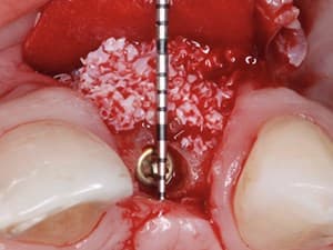
Immediate Dentoalveolar Restoration - Labial Contour Grafting at the Time of Immediate Placement
Description:
Until recently, tooth replacement at compromised sites in the aesthetic zone, required long-term treatment with possible undesirable complications in the tissue architecture. The Immediate Dentoalveolar Restoration (IDR) is a technique established to broaden indications for immediate loading on individual teeth. In this way, tissue losses with varied extensions are reconstructed in the same surgical session of implant placement and provisional crown installation, reducing the number of interventions and keeping predictability on aesthetic aspects. The IDR, which advocates minimally invasive procedures, will be presented as a viable and reproducible alternative.
Date Added:
5/21/2015
Author(s):
José Carlos Martins da Rosa, DDS, MS
Dr. José Carlos Martins da Rosa received his DDS degree in 1988 from the Federal University of Santa Maria/RS - Brazil. In 1992 he got a post graduate certificate in Pe...
[read more]
|

|
Online Videos / Surgery / Bone Grafting / Immediate Dentoalveolar Restoration - Labial Contour Grafting at the Time of Immediate Placement
Questions & Comments
|
Bakhadir Usmanov - (3/18/2019 12:57 AM)
Great technique! thank you!
|
Mohamed fouda420 - (2/16/2018 7:44 PM)
fantastic technique .. but i need to know how to gain the tuberosity graft
|
Christopher Bonin - (8/25/2015 4:10 PM)
Very inspirational. Thanks for posting.
|
Xiaoxiao Cai - (8/14/2015 8:03 PM)
amazing
|
Maurice Salama - (7/8/2015 11:35 AM)
Outstanding and creative solution to a common dilemma. My compliments. regards Maurice
|
Edison Louie - (7/7/2015 12:15 AM)
Thank you for your excellent presentation. Your results are very impressive.
|
carlo poggio553 - (7/6/2015 12:19 PM)
Amazing clinician!
|
Rigel Escalante Aburto - (5/25/2015 10:10 PM)
Excelente técnica. Muchas gracias por compartir esto con nosotros Dr.Martins da Rosa. Donde puedo conseguir el instrumental para tomar el injerto? los cinceles?
|
Shawn Tokunaga - (5/24/2015 12:01 PM)
Really enjoyed your presentation and unique technique. It was elegant and simple in its logic. It takes me outside of the "box" and I can resist trying this out. Very creative utilization of readily available bone while minimizing surgical trauma to the patient. Thank you for sharing this on Dentalxp.
|
|
Related Videos |
|
|
|
A Defined Algorithm for Regenerative Success Part 1 of 2
These presentations will focus open the above concepts but prioritize the diagnostic phase and surgical common denominators required for a successful regenerative outcome. Flap design, space maintenance, bone and membrane selections and tension free closure provides the template for all the new age materials and technologies to be successful in clinical practice.
Presented By:: |
Maurice Salama, DMD |
Presentation Style: |
Video |
Community Rating: |
|
|
Watch Now>>
|
|
|
|
|
Related Courses |
|
|
Alternative & New Tendencies for Alveolar Reconstruction in Modern Implant Dentistry
Knowledge of bone biology and physiology are crucial for successful dento-alveolar reconstruction. The presentation will review the natural healing process of the bone and the sequence of events needed for proper regeneration.
This process will then be discussed and compare with the present materials and technologies available in the market including alloplastics and allogenic materials. The role of the Stem Cells and rh-BMP2 will be exposed as part of these new trends in alveolar reconstruction for modern implant dentistry. Multiple cases will be presented demonstrating the versatility and outcomes in different clinical situations based on biological, bio-mechanics, and implant aesthetic needs.
Presented By:: |
Alejandro Vivas-Rojo, DDS, MS;Jesus A. Gomez, DDS |
Presentation Style: |
Online Self-Study Course |
CE Hours: |
1 CEU (Continuing Education Credit) |
|
Watch Now>>
|
|
|
|
|
A GRAFT-LESS Approach to Implant Dentistry
Vertical bone augmentation techniques are more invasive and success rates often vary according to the surgical expertise of the clinician. There has been a trend over time towards minimally invasive treatment options in implant dentistry. This approach attempts to avoid more complicated procedures involving bone augmentation for implant placement. This presentation discusses a “graft less” treatment philosophy that emphasizes the use of less demanding augmentation techniques for the purpose of placing short implants in atrophic posterior sites.
Presented By:: |
Craig M Misch, DDS, MDS |
Presentation Style: |
Online Self-Study Course |
CE Hours: |
1 CEU (Continuing Education Unit) |
|
Watch Now>>
|
|
|
|
|
Advanced Bone Grafting Techniques: Part 1 - Autogenous Bone Blocks in the Reconstruction of the Atrophic Mandibular Ridge
This lecture will describe the use of autologous onlay block bone grafting for reconstruction of moderate to severe atrophic posterior mandibular alveolar ridge. This will include a discussion of the various options to this form of grafting, such as Short implants, Lingual positioning, nerve re-positioning, distraction osteogenesis, and GBR. The Mutli-Tier onlay Bone Graft Technique will be highlighted in this presentation. Additionally, a review of a recently submitted research paper of long term follow up of implants placed into autologous bone blocks harvested from the mandibular ramus, retromolar and sympheseal areas will be discussed.
A description of surgical donor harvest sites in the mandible will also be performed as well as post op healing of these sites. Additional time will be spent reviewing causes of failure with bone blocks and implants placed into these sites.
Presented By:: |
Devorah Schwartz-Arad, DMD, PhD |
Presentation Style: |
Online Self-Study Course |
CE Hours: |
1 CEU (Continuing Education Unit) |
|
Watch Now>>
|
|
|
|
Related Articles |
|
|
Socket Augmentation: Rationale and Technique
This article presents the rationale behind socket augmentation for future implant placement and describes a technique that has shown to not only facilitate tooth extraction with minimal damage to the surrounding anatomic structures, but also to improve alveolar bone quality and quantity.
Author(s): |
Hom-Lay Wang, DDS, MSD, PhD;Koichi Kiyonobu, DDS, PhD; Rodrigo F. Neiva, DDS |
|
View Article>>
|
|
|
|
|
Long Term Follow-Up of Dental Implants Placed in Autologous Onlay Bone Graft
The aim of this study was to evaluate the efficacy of autologous intraoral onlay bone grafting (OBG) in correlation
with long-term survival rates of dental implants placed in the augmented bone. A retrospective study was conducted on 214 patients who received a total of 633 dental implants
placed in 224 autologous intraoral block OBG augmentations, combined with Bio-Oss – mixed with platelet-rich plasma
(PRP) and covered by platelet-poor plasma (PPP) – as scaffold, with a follow-up time up to 137 months. We suggest that augmentation of severely atrophied jaw bone through the placement of horizontal and/or vertical intraoral OBGs in combination with Bio-Oss saturated with PRP and covered by PPP should be considered a reliable, safe, and very effective surgical technique for obtaining high bone graft survival rate and high long-term implant
survival rate.
Author(s): |
Devorah Schwartz-Arad, DMD, PhD;Ronen Ofec, DMD, MSc; Galit Eliyahu, PhD; Angela Ruban, PhD; Nir Sterer, DMD, PhD |
|
View Article>>
|
|
|
|
|
Immediate Implant Placement into Extraction Sockets with Labial Plate Dehiscence Defects: A Clinical Case Series
To measure the buccal plate reconstruction of extraction sockets with labial plate dehiscence defects using a bone
allograft in combination with an absorbable collagen membrane and a custom-healing abutment at the time of tooth
removal. Implants placed into sockets with labial plate dehiscence defects demonstrated radiographic reformation of the labial plate dehiscence defect at 6 to 9 months post-treatment. The net gain in labial plate on CBCT in L1 and L2 was 3.0 mm, where 0 mm existed at pre-treatment. The minimum amount of labial plate thickness of 2.0 mm was achieved in all treated sites, evaluated radiographically at 6-9 months post-operatively, in a single
procedure, without flap elevation and maintaining the gingival architecture and satisfactory esthetics.
Author(s): |
Stephen J. Chu, DMD, MSD, CDT;Dennis P. Tarnow, DDS;Guido O. Sarnachiaro, DDS; Evangelina Sarnachiaro, DDS; Sergio Luis Gotta, DDS |
|
View Article>>
|
|
|
|
|