Login
Register
Login
Register
UPCOMING WEBINARS
View All Webinars
Webinar Presenter:
Robert Silva,Webinar Date: April 18, 2024
CE Hours: 1
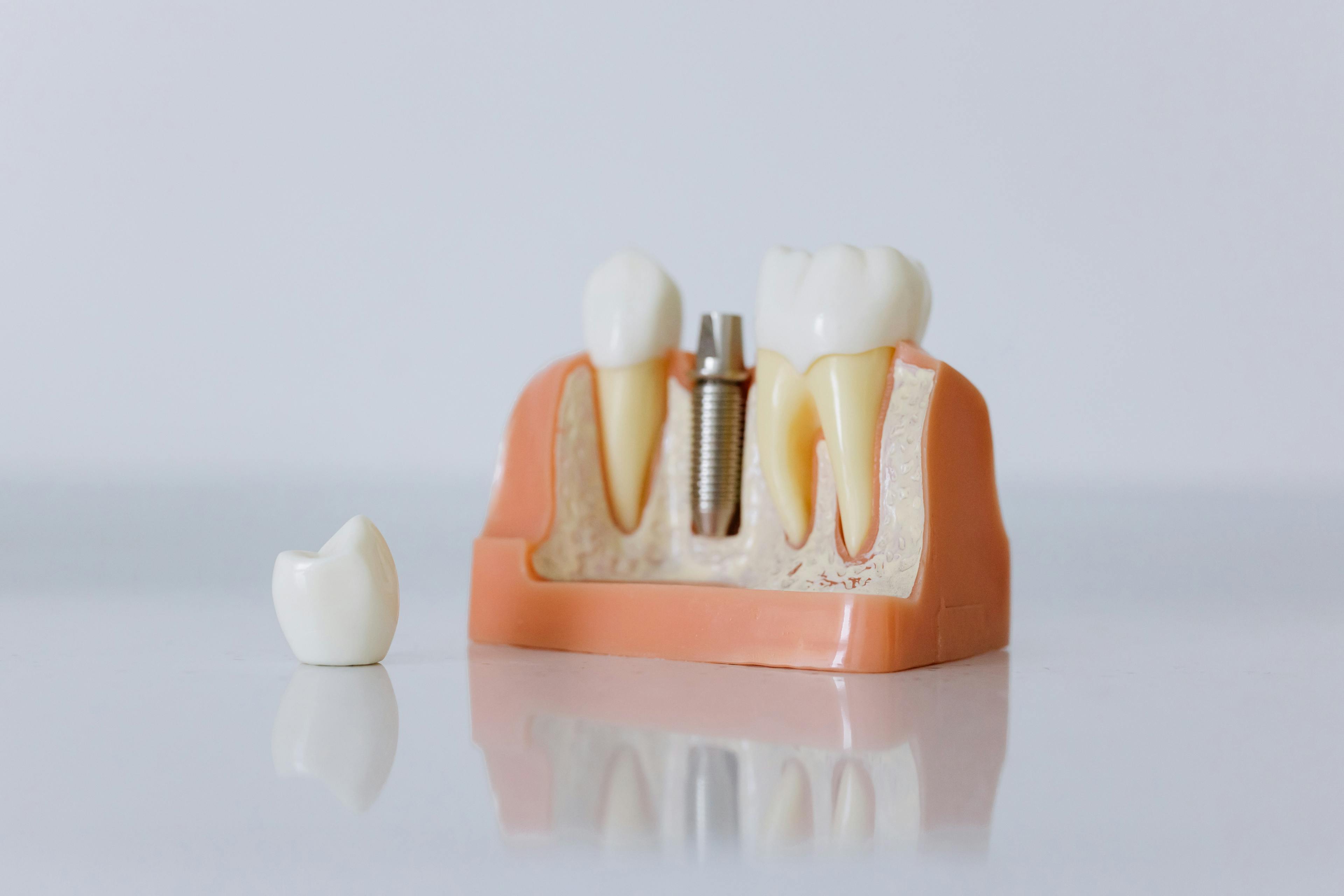
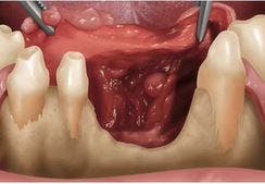
Proper flap management is critical and key to obtain tension-free flap closure at large 3D ridge augmentation sites. The objective of this presentation is to hi...
View Webinar
FROM THE FORUM
View DXP Forum
LIVE EVENTS
View All Events
NEW CONTENT
View All Content
- 3/21/2024
IV Sedation and When to Introduce an Anesthesiologist
- 3/18/2024
This course will show oral rehabilitation of a young female patient, following protocol in digital dentistry, which includes 4 phases.
OUR CONTINUING DENTAL EDUCATION SPECIALTIES
TRY BEFORE YOU BUY
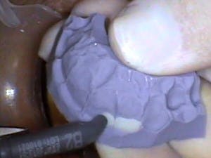
Using a Flexitime® Matrix in Anterior Composite Restorations
Feb 17, 2010
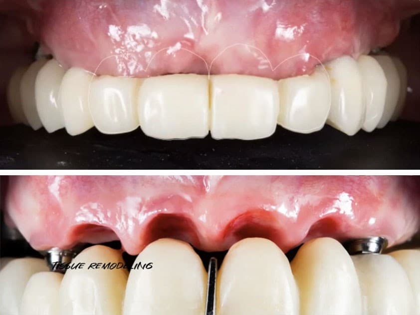
Proof of Principle: human histologic demonstration of socket healing with socket shield and grafting using non-demineralized autologous tooth dentin graft
Apr 24, 2023
The Replacement of Small-Diameter Teeth in the Esthetic Zone Using Narrow-Diameter Implants
Sep 20, 2022
4 Steps to a Predictable Full Arch Rehabilitation
Jun 1, 2021
ATTEND EVENTS WORLDWIDE

Date: 5/16/2024
Location: Modena, Italy


.jpg&w=3840&q=75)