Video Details
|
新時代の審美:組織の再構築と補綴とセラミック
Description:
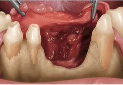
This presentation will focus on ideal implant placement through maintaining the bucco-lingual dimension of bone following extraction through extraction socket preservation techniques. The application of bone regenerative materials and soft tissue surgical preservation techniques are essential to an esthetic result and will be discussed at length. A review of the literature and useful clinical "tricks" techniques in combination with the hands on portion of the presentation will allow you to be comfortable with bone-grafting of extraction sockets for future implant placement.
Date Added:
12/20/2011
Author(s):
Mario A. Vilardi, DMD
Dr. Mario A. Vilardi received his DMD from the University of Pennsylvania School of Dental Medicine in 1974, where he later was awarded a teaching fellowship where he p...
[read more]
|

|
Online Videos / Surgery / Bone Grafting / Optimum Implant Esthetics Through Extraction Socket Preservation Techniques.
Questions & Comments
|
Joon Kim554 - (7/5/2017 3:42 AM)
Hi, I enjoyed your presentation.
I would like to ask you about the difference between the internal barrier vs. external barrier membrane techniques.
I personally would like to visualize buccal defect and elevate a flap. I think it helps degranulate or currettage the socket easier.
And if the entire buccal wall is gone, internal barrier may not work unless it's OK to extend it all the way to mesial and distal wall to hold the membrane.
I think that there is less chance of graft material leaving the socket for internal barrier. What do you think?
|
Jose Abitbol - (8/9/2016 12:17 PM)
Very good lecture! Thank you!
|
Jose Abitbol - (8/9/2016 12:17 PM)
Very good lecture! Thank you!
|
Jose Abitbol - (8/9/2016 12:16 PM)
Very good lecture! Thank you!
|
Jose Abitbol - (8/9/2016 12:16 PM)
Very good lecture! Thank you!
|
Tyler Binner - (12/30/2011 10:19 AM)
Nice presentation, thank you
|
mario vilardi - (12/20/2011 6:10 PM)
You don't actually delay healing by using a slowly resorbing bone graft, the bone heals around the particles at the same rate just the scaffold remains a lot longer. My studies of extraction socket repair showed less shrinkage by using a slow resorbing bone graft when compared to allograf materials.
|
mario vilardi - (12/20/2011 3:30 PM)
Dear Robert Klugman,
The case did not go back and forth but rather I substituted a different final case to show the way I am now closing or sealing the BioOss Collagen Bone grafting material, Sorry if that misled you, I should have made that clear.
|
mario vilardi - (12/20/2011 3:26 PM)
The bone grafting material was covered by Atrisorb Free Flow made by Zila
|
|
Related Videos |
|
|
|
Modern Implant Dentistry: Rules of Engagement in the Esthetic Zone - Part 1 of 2
This presentation will focus on the interdisciplinary relationship of the restorative dentist, periodontist and orthodontist to reconstruct the soft tissue foundation for all of these restorative options in anterior tooth replacement. The diagnosis of deficiencies as well as the varied treatment options will be discussed in detail. This includes periodontal crown lengthening, esthetic periodontal plastic soft tissue grafting procedures as well as prescription adjunctive orthodontic tooth movement to manipulate the soft tissue foundation prior to or subsequent with the restorative options of implants, bridges, or pontic replacement.
Presented By:: |
Maurice Salama, DMD;David Garber, DMD |
Presentation Style: |
Video |
Community Rating: |
|
|
Watch Now>>
|
|
|
|
|
Related Courses |
|
|
|
CadCam Customized 3D-Printed Subperiosteal Implant
When considering the future of implants, it appears justified to reconsider the old concepts in light of the availability of contemporary technologies, such as computer-aided design, softwares and programs design, virtual stress– strain testing, and 3D printing of titanium alloy. With the employment of these modern technologies, the concept of a “high-tech” subperiosteal implant has gradually emerged. Biological design of subperiosteal implant is mandatory for the success of the treatment, guidelines on architectural changes, topological optimization, and biofunctionalization will be explained. This new era of a “high-tech” customized 3D printed subperiosteal implant leads us to consider this technique s a valuable treatment option for atrophic jaws, avoiding more invasive procedures and giving the possibility of immediate loading, improving the patient’s quality of life.
Presented By:: |
Vladimir Garcia Lozada, DDS, MS, PhD |
Presentation Style: |
Online Self-Study Course |
CE Hours: |
1 CEU (Continuing Education Unit) |
|
Watch Now>>
|
|
|
|
|
Modern Implant Dentistry: Rules of Engagement in the Esthetic Zone - Part 1 of 2
This presentation will focus on the interdisciplinary relationship of the restorative dentist, periodontist and orthodontist to reconstruct the soft tissue foundation for all of these restorative options in anterior tooth replacement. The diagnosis of deficiencies as well as the varied treatment options will be discussed in detail. This includes periodontal crown lengthening, esthetic periodontal plastic soft tissue grafting procedures as well as prescription adjunctive orthodontic tooth movement to manipulate the soft tissue foundation prior to or subsequent with the restorative options of implants, bridges, or pontic replacement.
Presented By:: |
Maurice Salama, DMD;David Garber, DMD |
Presentation Style: |
Online Self-Study Course |
CE Hours: |
1 Continuing Education Unit (CEU) |
|
Watch Now>>
|
|
|
|
|
Biological Bone Augmentation- Only for REAL BONE BUILDERS
This webinar is addressed to all clinicians, who want to learn safe and predictable bone harvesting and augmentation techniques for different clinical situations. Clear guidelines for the use of augmentative procedures like the Split bone technique by Prof. Khoury and Biological Bone Augmentation with pure autogenous bone and advanced soft tissue management in order to achieve predicable results will be provided.
Presented By:: |
Frank Zastrow, DDS, M.Sc. |
Presentation Style: |
Online Self-Study Course |
CE Hours: |
1 CEU (Continuing Education Unit) |
|
Watch Now>>
|
|
|
|
Related Articles |
|
|
Socket Gafting with the use of Autologous Bone - an Experimental Study in the Dog
In five beagle dogs, the distal roots of the third and fourth mandibular premolars were removed. The sockets in the right or the left jaw quadrant were grafted with either anorganic bovine bone or with chips of autologous bone harvested from the buccal bone plate. After 3 months of healing, biopsies of the experimental sites were sampled, prepared for buccal–lingual ground sections and examined with respect to size and composition.
|
|
|
|
Subclassification and Clinical Management of Extraction Sockets with Labial Dentoalveolar Dehiscence Defects
Immediate implant therapy involving implants placed into intact Type 1 extraction sockets has become a consistent clinical technique. The classification of Type 2 extraction sockets, where the mucosal tissues are present but there is a midfacial osseous dehiscence defect, has been described according to the extent of the buccal bone plate absence. The literature has offered different techniques in the treatment of Type 2 sockets; however, the extent of the defect has never been defined or delineated.
Author(s): |
Stephen J. Chu, DMD, MSD, CDT;Dennis P. Tarnow, DDS;Guido O. Sarnachiaro, DDS; Mark N. Hochman, DDS |
|
View Article>>
|
|
|
|
|
Flapless Postextraction Socket Implant Placement in the Esthetic Zone: Part 1. The Effect of Bone Grafting and/or Provisional Restoration on Facial-Palatal Ridge Dimensional Change
The dental literature has reportted vertical soft tissue changes that can occur with immediate implant placement, bone grafting, and provisional restoration ranging from a gain or loss of 1.0mm. However, little is known of the effects of facial-palatal collapse of the ridge due to these clinical procedures. Based upon treatment modalities rendered, an ensuing contour change can occur with significant negative esthetic consequences. The results of a retrospective clinical cohort study evaluating the change in horizontal ridge dimension associated with implant placement in anterior postextraction sockets are presented for four treatment groups. Therefore, it is recommended to place a bone graft and contoured healing abutment or provisional restoration at the time of flapless postextraction socket implant placement.
Author(s): |
Dennis P. Tarnow, DDS;Stephen J. Chu, DMD, MSD, CDT;Maurice Salama, DMD;Christian F.J. Stappert, DDS, MS, PhD, Priv-Doz;Henry Salama, DMD;David Garber, DMD;Guido O. Sarnachiaro, DDS; Evangelina Sarnachiaro, DDS; Sergio Luis Gotta, DDS; Hanae Saito, DDS, MS |
|
View Article>>
|
|
|
|
|