Video Details
|
リッジスプリッティングテクニックにおけるピエゾサージェリーの役割 Part2
Description:
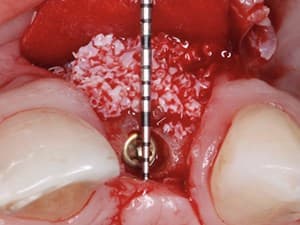
In this 2nd of a series, Dr. Dan Holtzclaw demonstrates the role of piezosurgical mediated ridge split technique in the reconstruction of ridge width deficiencies in order to facilitate more optimal placement of dental implants. A number of different variations exist regarding this technique. However, in this section, Dr. Holtzclaw discusses the role of piezoelectric hinge assisted ridge split (PHARS) technique in creating a hinged ridge split that is filled with particulate allograft and covered with a barrier membrane. Specific emphasis is placed on outlining the advantages and unique challenges of the PHARS technique.
Date Added:
7/4/2011
Author(s):
Dan Holtzclaw, DDS, MS
Dr. Holtzclaw received his dental degree (Cum Laude) from the University of Texas Health Science Center at San Antonio. During a subsequent 12 year ...
[read more]
|

|
Online Videos / Surgery / Bone Grafting / The Role of Piezo-Surgical Ridge Split Technique in the Reconstruction of Ridge Deficiencies - Part 2 of 2
Questions & Comments
|
Amr Azim - (4/8/2021 6:58 AM)
Excellent presentation, I have went through it several times in the last 3 years while preparing my lectures around the subject. have you started considering ridge expansion and simultaneous osseo-densification as an alternative in some cases, I mean using the Densah concept.
|
Juan Uribe Saldarriaga - (3/10/2016 2:49 PM)
Very nice presentation congratulations Dr Dan, I just want to know, what are you doing now when you need to gain height and what bone regeneration procedures in vertical case you do. Thanks
|
henry salama - (10/10/2015 9:27 AM)
Harry, since the idea is to "split" (not just cut through) the buccal cortical plate from the palatal/lingual bone plate, we would require an intervening layer of cancellous bone. Therefore, the minimum thickness that can be "split" is likely to be about 3mm, i.e. 1mm buccal, 1mm cancellous and 1mm palatal. Also, Harry, why not post images of the CT in question on the Forum. I'm sure you will get a great deal of feedback. Good luck.
|
Harry Albers - (10/9/2015 4:56 PM)
What is the minimum thickness of a ride to due a ridge split technique? Can I send you a cone beam of a case?
Harry Albers
|
byron scott - (5/26/2014 12:16 PM)
Very informative & straightforward.
|
Eviatar Buchman - (10/24/2011 5:32 PM)
Thank you very much for your presentation. In a case of braking the buccal wall how do you recomand to fix it. Is there any video about it. second question, what if the risk to damage the inferior nerve?
E. Buchman
|
dentimp huang - (7/23/2011 2:11 PM)
Great answer ! Thank you , Dr.Holtzclaw.
|
dan holtzclaw - (7/19/2011 5:03 PM)
Thank you for the kind comments.
To answer everyone's questions:
1) I generally will not place implants simultaneously in the mandible. The bone here is very dense, especially with a resorbed ridge. That is why I employ the apical hinge cut. This allows me to lateralize the buccal plate more extensively. With this being said, however, it also makes the area less stable for simultaneous implant placement. In the maxilla, I generally have no problem with placing implants with a ridge split (circumstances permitting)
2) I have only used PRF and have not used PRGF. They both have their supporters. It is very fun to watch Chokroun and Anitua square off against one another. I have seen great cases from both sides.
3) I have found that the ridge heals just fine with a full thickness flap. I understand the concept for a split thickness flap, but have not found it to be necessary.
|
dan holtzclaw - (7/19/2011 4:58 PM)
test
|
|
Related Videos |
|
|
|
|
Maximizing Aesthetics on Adjacent Implants and Full Arch Reconstructions - Part 2 of 2
The beauty in implant dentistry is that every case is an individual challenge. An ideal implant treatment uses procedures , techniques, materials and technologies that leads fast and on the shortest way to a maximally aesthetic result. This is our goal, in every clinical case, from single tooth replacement to full arch reconstructions.
Presented By:: |
Henriette Lerner, DMD |
Presentation Style: |
Video |
Community Rating: |
|
|
Watch Now>>
|
|
|
|
|
CadCam Customized 3D-Printed Subperiosteal Implant
The “classical” subperiosteal implant does not enjoy a good reputation; the causes of failure were myriad, as evidenced in the literature. When considering the future of implants, it appears justified to reconsider the old concepts in light of the availability of contemporary technologies, such as computer-aided design, softwares and programs design, virtual stress– strain testing, and 3D printing of titanium alloy. With the employment of these modern technologies, the concept of a “high-tech” subperiosteal implant has gradually emerged. This new era of a “high-tech” customized 3D printed subperiosteal implant leads us to consider this technique a valuable treatment option for atrophic jaws, avoiding more invasive procedures and giving the possibility of immediate loading, improving the patient’s quality of life.
Presented By:: |
Vladimir Garcia Lozada, DDS, MS, PhD |
Presentation Style: |
Video |
Community Rating: |
|
|
Watch Now>>
|
|
|
|
Related Courses |
|
|
Current Concepts in Bioactivity and Regeneration
In this lecture, tips on how to deal with the different types of extraction socket, mostly infected, will be presented. The use of plasma rich in growth factors and the Endoret® (PRGF®) fibrin membrane is a key factor to enhance socket regeneration, post-operative recovery and minimize/treat complications.
Presented By:: |
Eduardo Anitua, MD, DDS, PhD |
Presentation Style: |
Online Self-Study Course |
CE Hours: |
1 CEU (Continuing Education Unit) |
|
Watch Now>>
|
|
|
|
|
A GRAFT-LESS Approach to Implant Dentistry
Vertical bone augmentation techniques are more invasive and success rates often vary according to the surgical expertise of the clinician. There has been a trend over time towards minimally invasive treatment options in implant dentistry. This approach attempts to avoid more complicated procedures involving bone augmentation for implant placement. This presentation discusses a “graft less” treatment philosophy that emphasizes the use of less demanding augmentation techniques for the purpose of placing short implants in atrophic posterior sites.
Presented By:: |
Craig M Misch, DDS, MDS |
Presentation Style: |
Online Self-Study Course |
CE Hours: |
1 CEU (Continuing Education Unit) |
|
Watch Now>>
|
|
|
|
|
Achieving Stable Esthetic Results with Implant Supported Restorations
We will discuss basic concept regarding immediate implant placement and guided bone regeneration procedure related to the esthetic zone before focusing on the soft tissue management. We will describe the prosthetic procedures which are performed before, during and after the surgical procedures. Provisional restorations, impression techniques, prosthetic profiles and restorative materials will be presented. The purpose of the presentation is to provide a check list that will guide the clinician developing a proper analysis and diagnosis for the successful esthetic result with implant supported restoration.
Presented By:: |
Gianluca Paniz, DDS, MS, FACP;Luca Gobbato, DDS, MS |
Presentation Style: |
Online Self-Study Course |
CE Hours: |
1 CEU (Continuing Education Unit) |
|
Watch Now>>
|
|
|
|
Related Articles |
|
|
Socket Gafting with the use of Autologous Bone - an Experimental Study in the Dog
In five beagle dogs, the distal roots of the third and fourth mandibular premolars were removed. The sockets in the right or the left jaw quadrant were grafted with either anorganic bovine bone or with chips of autologous bone harvested from the buccal bone plate. After 3 months of healing, biopsies of the experimental sites were sampled, prepared for buccal–lingual ground sections and examined with respect to size and composition.
|
|
|
|
A Fixed Whole-Mouth Rehabilitation Utilizing Natural Abutments and Implants: Treatment Concepts and Clinical Realization
A 45-year-old female patient presented to the clinic with a request to treat her deteriorating dentition that had been reconstructed 15 years ago with fixed restorations. Clinical examination revealed fixed partial dentures cemented to natural abutments in the maxilla, whereas telescopic restorations were cemented to natural abutments bilaterally in the mandible. The treatment plan included a whole-mouth rehabilitation utilizing natural teeth and implants. As the patient declined any surgical augmentation…
Author(s): |
Prof. Nitzan Bichacho, DMD;Rafi Lahav, MDT, Cobi J. Landsberg, DMD |
|
View Article>>
|
|
|
|
|
|