Video Details
|
根管穿孔の診断と治療 Part3
Description:
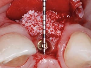
In this dynamic video presentation, Dr. Leandro Pereira details the diagnosis and variable options for the management and treatment of root perforations. The advantages of CBCT in the diagnostic and evaluation phase as well as surgical microscopy in the treatment phase are discussed. A thorough algorithm that includes location, size and shape of the perforation is presented to guide the clinician in assessing the potential for success in clinical intervention. Materials as well as surgical vs. non-surgical treatments are also discussed.
Date Added:
5/6/2012
Author(s):
Leandro A. P. Pereira, DDS, MSc
Dr. Leandro A. P. Pereira received his DMD from the Universidade Estadual de Campinas, UNICAMP of Piracicaba Dental School FOP in 1997. In 2000 he received his specialty certi...
[read more]
|

|
Online Videos / General / Endodontics / Root Canal Perforations - Diagnosis and Treatment - Part 3 of 3
Questions & Comments
|
Marcel Fundora - (8/12/2020 8:38 PM)
great presentation, thank you!. new graduate question: if CaSO4 is placed at the surgical site to prevent CT ingrowth, what happens when it dissolves after 4 weeks? would CT not invaginate at that point considering bone tissue takes longer to fill in? would you consider an allograft instead? thank you!
|
Eric Pena - (11/27/2016 6:53 PM)
Thank you! Great presentation.
|
Leandro Pereira56 - (5/20/2012 7:02 PM)
Dear Dimitris, the white line is a thin metallic line inside this Angelus Fiber Post. This line helps to visualize the fiber post extension . It's just an Xray contrast.
|
Dimitris Tsanaktsidis - (5/13/2012 3:50 PM)
Well done Leonardo.Very nice presentation in all parts. I would like to ask you what is that thin white line that is in all x-rays on the lateral incisor?
|
Glen Carmo15 - (5/1/2012 7:11 PM)
Wowwwww, You did again....congrats Leandro
|
Glen Carmo15 - (5/1/2012 7:11 PM)
Wowwwww, You did again....congrats Leandro
|
Related Videos |
|
|
|
|
Implant Dentistry: The Team Approach
In this lecture Dr Stanley will present his "No Half Smiles” Philosophy and how an effective treatment plan and its correct team approach can help save time and stress. For over a decade Dr Stanley and is interdisciplinary team have been working together as a tight unit on complex cases. From the old school approach to the latest DSD surgical planning, with combined Endodontics, Orthodontics and cosmetic implant dentistry, the cases presented will show how his team approaches each individual case for optimal outcome.
In this lecture a lot of “behind the scenes” explanations will be given on the why, how and what his team do to get consistent excellent results and manage a stress free environment in his practice.
Presented By:: |
Miguel Stanley, DDS |
Presentation Style: |
Video |
Community Rating: |
|
|
Watch Now>>
|
|
|
|
|
|
CBCT in Endodontics: Changing the Landscape of Diagnosis and Clinical Treatment
Radiographic imaging is essential in the diagnosis, treatment planning and follow-up in endodontics. The interpretation of an image can be confounded by a number of factors, including the regional anatomy as well as superimposition of both the teeth and surrounding dento-alveolar structures. As a result of superimposition, periapical radiographs reveal only limited aspects, a 2-dimensional view, of the true three dimensional anatomy. Additionally, there is often geometric distortion of the anatomical structures being imaged with conventional radiographic methods. These problems can be overcome by utilizing small or limited volume cone beam computed tomography (CBCT) imaging techniques which produce accurate 3-dimensional images of the teeth and surrounding dento-alveolar structures. This presentation will highlight the indications, advantages and considerations of the use of CBCT in endodontics.
Presented By:: |
Frederic Barnett, DMD |
Presentation Style: |
Video |
Community Rating: |
|
|
Watch Now>>
|
|
|
|
Related Courses |
|
|
Mastering Anterior Tooth Fracture with Contemporary Composites: The i3D Layering Technique
The aim of this presentation is to present an individual three-dimensional layering technique with contemporary composites. This novel procedure is indicated in moderate to extensive crown fractures and is considered predictable after eliminating the parameter of thickness while layering restorative materials.
Presented By:: |
Yiannis P. Brokos, DDS, MSc, Clinical Specialty in Operative Dentistry |
Presentation Style: |
Online Self-Study Course |
CE Hours: |
1 CEU (Continuing Education Unit) |
|
Watch Now>>
|
|
|
|
|
Optimized Instrumentation & Technology in Endodontic Therapy
Rotary instrumentation using Optimized Torque Reduction (OTR) motion and Apex Locator-Handpiece technology has the potential to improve clinical efficiency during endodontic therapy. In this webinar, Dr. Nasseh explains the implementation of one such technology using the ESX instrumentation and obturation system. He further shares some tips and settings for effective, safe operations. Various endodontic motions and their indications for use are explained and an anterior tooth demonstration is presented to consolidate all covered points with a clinical demonstration.
Presented By:: |
Allen Ali Nasseh, DDS, MMSc |
Presentation Style: |
Online Self-Study Course |
CE Hours: |
1 CEU (Continuing Education Unit) |
|
Watch Now>>
|
|
|
|
|
Prevention and Management of Broken Endodontic Instruments
This lecture aims to review the most common preventive measures to avoid file breakage and to provide clinical tips and recommendations for the management of challenging broken file cases. Highly invasive, minimal invasive and non invasive broken instrument removal techniques are showcased through video case presentation series under the microscope.
Presented By:: |
Antonis Chaniotis, DDS MDSC |
Presentation Style: |
Online Self-Study Course |
CE Hours: |
1 CEU (Continuing Education Unit) |
|
Watch Now>>
|
|
|
|
Related Articles |
|
|
Endodontic-Restorative Symbioses: Diagnosis and Treatment
The success of endodontic treatment is comprised of proper diagnosis and treatment, which is easier to accomplish when the symptoms correlate to the clinical and radiographic findings. It is generally a more predictable treatment on anterior teeth as opposed to posterior teeth due to the number of canals and their internal configuration. A multidisciplinary approach has never been more essential to provide the patient with the best possible choices for treatment. Moreover, the general dentist and…
|
|
|
|
Do All Endodontically Treated Teeth Require a Post?
Treatment modalities in restorative dentistry should be implemented with a solid rationale behind them and support from scientific studies.These principles, in conjunction with clinical experience, can dictate when and if a post is required when a tooth has undergone root canal treatment. Dentists know each tooth is anatomically different and should be diagnosed and treated with an individual approach when it comes to its restoration. It is tempting to follow a path that leads into a routine in which…
|
|
|
|
Dental Endoscopic Viewing System
Endoscopy has enabled medicine to take a quantum leap in disease diagnosis and therapeutic management by allowing visualization and access for repair of injuries via minimally invasive surgical (MIS) techniques. Since their introduction, fiberoptic bundles and electronic video technology have continually advanced and fostered smaller-bore (viewing tubes) endoscopes of 2 mm and less, further improving access into smaller cavities and anatomical spaces of the body. This viewing technology has reached…
Author(s): |
Ronald Goldstein, DDS;David Garber, DMD;Maurice Salama, DMD;Henry Salama, DMD;Lee H. Silverstein, DDS, MS, FACD, FICD |
|
View Article>>
|
|
|
|
|