Video Details
|
Selection of Biomaterials, Bone Grafts and Growth Factors in Implant Therapy
Description:
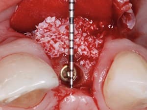
In this highly evidence based and detailed technique presentation, Dr. Daniel Buser discusses decision making and surgical protocols in the placement of implants in compromised anterior sites. A focus is also placed on selection of biomaterials, bone grafts and growth factors.
Date Added:
7/3/2017
Author(s):
Daniel Buser, DDS
Dr. Daniel Buser is Professor and Chairman at the Department of Oral Surgery at the University of Bern in Switzerland. He spent 3-times a sabbat...
[read more]
|

|
Online Videos / Surgery / Bone Grafting / Selection of Biomaterials, Bone Grafts and Growth Factors in Implant Therapy
Questions & Comments
|
Maurice Salama - (11/9/2017 3:20 PM)
Back action chisels or bone harvest scrapers....regards Dr. Salama
|
kevin potocsky - (11/9/2017 12:26 PM)
what instrument is best for scraping this autogenous bone??
|
George Duello - (7/12/2017 2:22 PM)
Dear Dr. Buser,
Excellent presentation support your findings and papers on the single teeth treatments with Type II cases in the exthectic zone. Thanks for sharing on paper.
I thank you and DentalXP for showing your findings-it will be a new part of my options for patients.
George Duello-DentalXp expert.
|
Maurice Salama - (7/2/2017 7:51 PM)
Loan and Scott; Professor Buser and Urs Belser will be the KEYNOTE speakers at the next Dentalxp Global Symposium in South Florida Feb. 8-11th 2018.
|
Scott Ly - (7/2/2017 6:59 PM)
Loan, his courses are at the university of Bern in Switzerland
|
FİGEN ÖZÇELEBİ - (11/4/2016 9:16 AM)
Thank you for your wonderful presentation Dr Buser
|
Long Nguyen - (10/15/2016 12:10 PM)
Thank you so much for a wonderful wonderful presentatiion. We learn a lot from you. Very helpful. Lookiing forward to more and more lectures and hand- on from you. How could we attend your seminars?
Thanks DentalXP posting this on for us.
|
Maurice Salama - (10/14/2016 7:00 AM)
A very effective protocol for GBR and tooth replacement. If you can follow these rules it will make you a better implant dentist for sure. Dr. Salama
|
aryeh gellman - (10/14/2016 3:18 AM)
great methods displayed and lecture too
tnx
|
|
Related Videos |
|
|
Timing of Implant Placement After Extraction in the Esthetic Zone
In this presentation, Dr. Daniel Buser outlines the most current scientific knowledge concerning the clinically relevant challenge of when and how to place implants after extraction of a tooth in the esthetic zone. He also gives an update on treatment options and tissue biology.
Presented By:: |
Daniel Buser, DDS |
Presentation Style: |
Video |
Community Rating: |
|
|
Watch Now>>
|
|
|
|
|
|
Related Courses |
|
|
Bone Augmentation & Vascular Enhancement Concepts: It’s Time to Make it Simple!
Growing bone is fascinating, and clinicians have developed many techniques in that matter, most of them restricted to an elite. Yes, it does take a lot of skills to harvest bone blocks, to stabilize them with screws etc, and without forgetting the soft tissue handling as it can be a real challenge to cover the bone graft especially for a vertical augmentation.
The purpose of this presentation is to make the participant think biology first and not mechanic first. A biological thinking will dramatically simplify vertical and/or horizontal bone augmentation procedures. We will also be introducing a new approach for the simplification of soft tissue handling: the Soft Brushing Technique, that will allow the coronal advancement of both buccal and lingual flaps, but also the palatal flap without the need for periosteal releasing incisions.
Presented By:: |
Jérôme Surmenian, DDS |
Presentation Style: |
Online Self-Study Course |
CE Hours: |
1 CEU (Contining Education Unit) |
|
Watch Now>>
|
|
|
|
|
Complete Management of the Implant Site: From Simple Cases to Complex Situations
The health and the structure of the peri-implant tissue is vital for a long term prognosis of aesthetic and functional dental treatments. The prevalence of peri-implant diseases and recessions is a problem that must be overcome by a perfect treatment planning, along with a step-by-step design of the future restorations. When the clinician places an implant, he must consider the 3D positioning regarding the hard and soft tissue. When one decide to restore an implant, he must be aware of the gingival volume and it’s position. In this presentation, we want to show fully documented cases, from bone grafting, soft tissue enhancement, to final restorations.
Presented By:: |
Mihnea Cafadaru, DDS |
Presentation Style: |
Online Self-Study Course |
CE Hours: |
1 CEU (Continuing Education Unit) |
|
Watch Now>>
|
|
|
|
|
Advanced Bone Grafting Techniques: Part 2 - Autogenous Bone Blocks in the Reconstruction of the Atrophic Maxillary Ridge
This lecture will describe the use of autologous onlay block bone grafting for reconstruction of moderate to severe atrophic maxillary alveolar ridge. This will include a discussion of the various options to this form of grafting, such as short implants, maxillary sinus augmentation, sub-nasal elevation procedure, connective tissue grafting and soft tissue manipulation, Le-Fort I down fracture/osteotomy and the use of progenitor cells: bone marrow aspirating concentrated.
A description of surgical donor harvest sites in the mandible will also be performed as well as post-op healing of these sites. Additional time will be spent reviewing causes of failure with bone blocks and implants placed into these sites.
Presented By:: |
Devorah Schwartz-Arad, DMD, PhD |
Presentation Style: |
Online Self-Study Course |
CE Hours: |
1 CEU (Continuing Education Unit) |
|
Watch Now>>
|
|
|
|
Related Articles |
|
|
Intraoral Autogenous Block Onlay Bone Grafting for ExtensiveReconstruction of Atrophic Maxillary Alveolar Ridges
Autologous bone grafting used with dental implants was originally described by Bränemarketal in 1975, and is now a well-accepted procedure in oral and maxillofacial rehabilitation.Placement of an endosseous implant requires sufficient bone volume for complete bone coverage. Furthermore, the pattern of ridge resorption contributes to an unfavorable maxillomandibular relationship, requires angulations of the implants and/or angled abutments, and affects the proximity of adjacent facial concavities(maxillary sinus, nasal cavity) and vital structures(mandibular nerve).
|
|
|
|
Alloplastic Bone Substitutes Around Dental Implants
The repair of osseous defects has been a goal in dentistry for many decades. Subsequently, within the last several years, there has been an emergence of a new class of materials in dentistry referred to as synthetic bone. Finding the ideal bone substitute material has been the goal of researchers for many years. In attempting to achieve this ideal material, dental practitioners have tried with varying degrees of success: autogenous and demineralized freeze-dried bone, allografts of plastic, carbon…
Author(s): |
Lee H. Silverstein, DDS, MS, FACD, FICD;Lee M. Whitesides, DMD, MMSC |
|
View Article>>
|
|
|
|
|
Reconstruction of a Horizontal and Vertical Bone defect using The Cortical Lamina Technique
For many years guided bone regeneration has been a challenge in oral and implant surgery. Many
techniques and devices have been used in trying to regenerate and reconstruct resorbed edentulous
ridges. The literature in the field is scarce due to the impossibility to perform RCT on such
lesions, therefore the judgement of the international community is based on case reports and
retrospective studies.
In the last ten years a new device has proved to be quite successful and predictable on top of
showing a very low morbidity, a membrane made of xenogeneic collagenated bone with features
that are very different from any other material used in the past.
This paper will show the application of the cortical lamina technique where a vertical and
horizontal defect was corrected and successfully restored.
|
|
|
|