Video Details
|
SGFBR(スクリューガイデットファーストボーンジェネレーション)での下顎の水平垂直骨造成
Description:
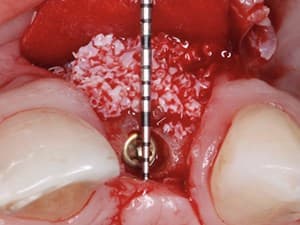
The region which often presents the greatest challenge for augmentation is the molar and premolar region of the Mandible. This is due to naturally occurring anatomical considerations. This presentation will highlight a less invasive surgical option to autogenous block transplantation and has displayed a very high success rate. The speed of bone regeneration with this technique provides for re-entry and implant placement after only 4 months.
Date Added:
2/14/2012
Author(s):
Roland Török, DMD
Dr. Roland Török is the founder of "Implant Institute Török" which was developed in 2008 to provide quality continuing education on the subject of dental implants. Since 2003 ...
[read more]
|

|
Online Videos / Surgery / Bone Grafting / SGFBR (Screw Guided Fast Bone Regeneration) Horizontal & Vertical Bone Augmentation of the Atrophic Mandible
Questions & Comments
|
Carlos Ramirez - (5/7/2014 12:16 AM)
Dr. Roland, I find this technique amazing, I really do. I saw the final prothetic picture and the 4 year after and I think I saw some resorption that perhaps compromised the papilla and tissue surrounding the prosthesis, maybe causing food impaction and difficult hygiene. So I would like to know, why do you prefer to go with the SF-GBR instead of doing S-GBR , wait 4 months instead of 2, and avoid the resorption in time?
Thanks.
|
Roland Toeroek Toeroek - (2/22/2012 9:48 AM)
@ Mario Marcone, if i can insert 1/3 of the implant in own bone and i have a wall wich goes to the coronal part of the implant i will make the technique what i show, if i have not enough own bone to have a good primar stability or at least one bone wall wich is in contact with my implant i will make first the SGFBR and after 4 moth insert the implant.
|
Roland Toeroek Toeroek - (2/22/2012 9:43 AM)
@ Paul Scholl, thank you too
|
Roland Toeroek Toeroek - (2/22/2012 9:43 AM)
@ Mark Evans, we used in every block augmentation case collagene membranes, my opinion is that the problem with the resorbtion is that we have a tension on the flap and we have a bad blood supply on the surface of the block witch is not in contact with the own bone
|
Roland Toeroek Toeroek - (2/22/2012 9:40 AM)
@ yasin demircan,we give the patients Amoxicilin 1000, 3 per day, one day before surgery, and 2 days after, after this for 5 more days 2 per day. We also give the patients Ibuprofen 1000, they should take it if they have pain.
|
Mario Marcone - (2/19/2012 1:45 PM)
Roland, You showed a case whereby you augmented a mandibular ridge using screws and implants as tenting devices at the same surgery. In which situations do you prefer to go with a staged technique using only screws on a first surgery ? In which situations do you elect to do a combined technique using both screws and implants simultaneously? Is it a matter of the amount of bone volume required ?
|
Paul Scholl - (2/19/2012 11:39 AM)
Beautiful cases and nice technique. Thanks for the information.
|
Mark Evans - (2/15/2012 1:27 PM)
DR Torok
Did these bone block examples include a covering membrane? My brief peruse of the literature suggests that substantial resorption rates are experienced if block are not covered by membrane.
Thanks you
Mark Evans
|
yasin demircan - (2/15/2012 5:46 AM)
excellent presentation.seems easy according to block grafts.my question is what is your medication to deal with infection,pain and mostly eudema?thank you?
|
|
Related Videos |
|
|
|
Maximizing Aesthetics on Adjacent Implants and Full Arch Reconstructions - Part 2 of 2
The beauty in implant dentistry is that every case is an individual challenge. An ideal implant treatment uses procedures , techniques, materials and technologies that leads fast and on the shortest way to a maximally aesthetic result. This is our goal, in every clinical case, from single tooth replacement to full arch reconstructions.
Presented By:: |
Henriette Lerner, DMD |
Presentation Style: |
Video |
Community Rating: |
|
|
Watch Now>>
|
|
|
|
|
Advanced Bone Grafting Techniques: Part 2 - Autogenous Bone Blocks in the Reconstruction of the Atrophic Maxillary Ridge
This lecture will describe the use of autologous onlay block bone grafting for reconstruction of moderate to severe atrophic maxillary alveolar ridge. This will include a discussion of the various options to this form of grafting, such as short implants, maxillary sinus augmentation, sub-nasal elevation procedure, connective tissue grafting and soft tissue manipulation, Le-Fort I down fracture/osteotomy and the use of progenitor cells: bone marrow aspirating concentrated.
Presented By:: |
Devorah Schwartz-Arad, DMD, PhD |
Presentation Style: |
Video |
Community Rating: |
|
|
Watch Now>>
|
|
|
|
Related Courses |
|
|
|
Advances in Bone Augmentation and Related Armamentarium
The ability to predictably and successfully augment bone volume is a critical surgical and technical requirement for any clinician involved in implant dentistry. This presentation outlines the evolution of diagnostic and therapeutic protocols in bone augmentation as well as introducing new and novel biologics and armamentarium being made available for clinicians to be more efficient and efficacious in this important endeavor.
Presented By:: |
Dr. Jan Kielhorn |
Presentation Style: |
Online Self-Study Course |
CE Hours: |
1 CEU (Continuing Education Unit) |
|
Watch Now>>
|
|
|
|
|
Bone Graft Cements: A New Advance to Augmentation Solutions
The purpose of this lecture is to shed light on the evolution of raw materials in order to find the ultimate bone graft cement, exploring the potential and the new opportunities of using cement-based augmentation materials in the maxillofacial and dental fields, and emphasizing the advantages, disadvantages and methods of use from the scientific and clinical points of view.
Presented By:: |
Amos Yahav, DMD |
Presentation Style: |
Online Self-Study Course |
CE Hours: |
1 CEU (Continuing Education Credit) |
|
Watch Now>>
|
|
|
|
|
Microsurgical Soft Tissue Reconstruction for Teeth & Implants - Part 1 of 2
This presentation is focused in the importance of soft tissue grafting techniques around teeth and implants and how to do it in a predictable, modern and a minimally invasive way.
We present many different techniques illustrated with clinical cases.
Presented By:: |
Ramon Gomez Meda, DDS |
Presentation Style: |
Online Self-Study Course |
CE Hours: |
1 CEU (Continuing Education Unit) |
|
Watch Now>>
|
|
|
|
Related Articles |
|
|
Alloplastic Bone Substitutes Around Dental Implants
The repair of osseous defects has been a goal in dentistry for many decades. Subsequently, within the last several years, there has been an emergence of a new class of materials in dentistry referred to as synthetic bone. Finding the ideal bone substitute material has been the goal of researchers for many years. In attempting to achieve this ideal material, dental practitioners have tried with varying degrees of success: autogenous and demineralized freeze-dried bone, allografts of plastic, carbon…
Author(s): |
Lee H. Silverstein, DDS, MS, FACD, FICD;Lee M. Whitesides, DMD, MMSC |
|
View Article>>
|
|
|
|
|
|
Immediate Implant Placement into Extraction Sockets with Labial Plate Dehiscence Defects: A Clinical Case Series
To measure the buccal plate reconstruction of extraction sockets with labial plate dehiscence defects using a bone
allograft in combination with an absorbable collagen membrane and a custom-healing abutment at the time of tooth
removal. Implants placed into sockets with labial plate dehiscence defects demonstrated radiographic reformation of the labial plate dehiscence defect at 6 to 9 months post-treatment. The net gain in labial plate on CBCT in L1 and L2 was 3.0 mm, where 0 mm existed at pre-treatment. The minimum amount of labial plate thickness of 2.0 mm was achieved in all treated sites, evaluated radiographically at 6-9 months post-operatively, in a single
procedure, without flap elevation and maintaining the gingival architecture and satisfactory esthetics.
Author(s): |
Stephen J. Chu, DMD, MSD, CDT;Dennis P. Tarnow, DDS;Guido O. Sarnachiaro, DDS; Evangelina Sarnachiaro, DDS; Sergio Luis Gotta, DDS |
|
View Article>>
|
|
|
|
|