Login
Register
Login
Register


Presented by:
Event Date(s): October 24 - 26, 2024
Location: Chantilly, VA, United States


Join us for the 9th edition of the ImplantoDays Congress 6 – 8 June 2024, in the breathtaking setting of Poiana Brașov.
Presented by: Howard Gluckman, BDS, MCHD, PHD, Christian Coachman, Maurice Salama, +5 moreMiguel Stanley, Marcelo Ferrer Balart, Henriette Lerner, Ventseslav Stankov, Roberto Carvalho da Silva
Affiliates: ImplantoDays
Event Date: June 6, 2024
View DetailsUPCOMING WEBINARS
View All Webinars
Webinar Presenter:
Rami Salloum,Webinar Date: May 8, 2024
CE Hours: 1
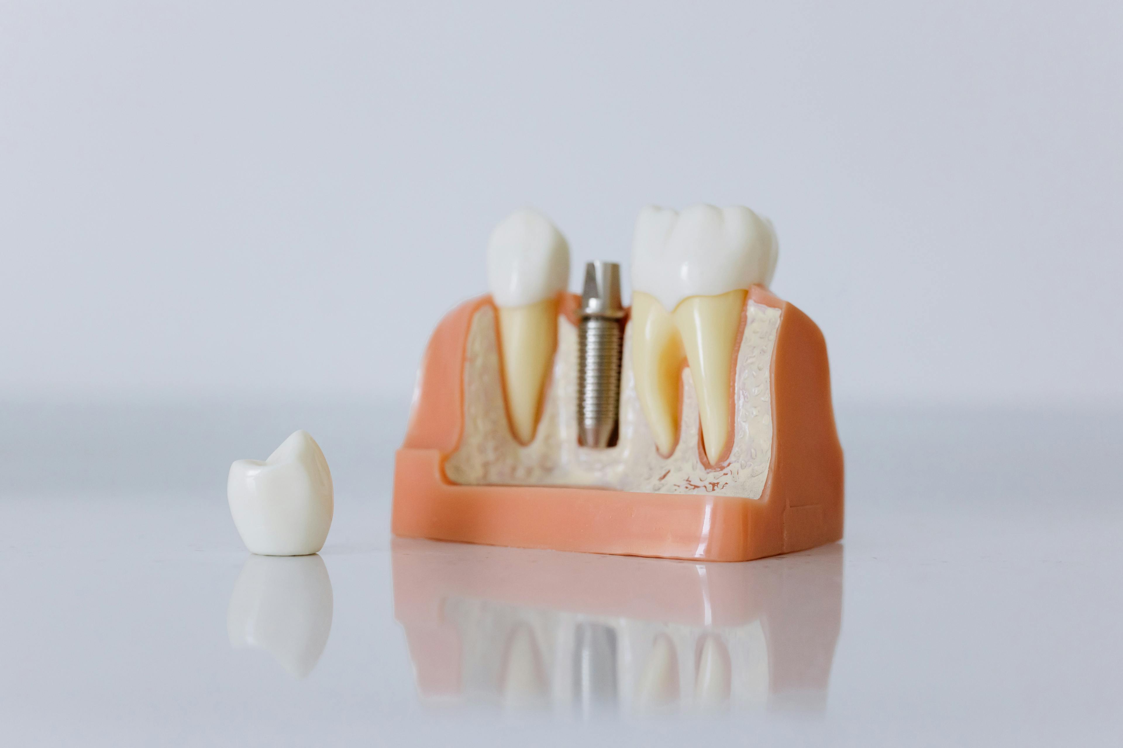
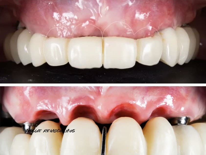
Proper treatment planning and guided implant placement will lead to an ideal prosthetic outcome. The accuracy of implant placement is crucial in a single too...
View Webinar
FROM THE FORUM
View DXP Forum
LIVE EVENTS
View All Events
NEW CONTENT
View All Content
- 5/6/2024
Proper treatment planning and guided implant placement will lead to an ideal prosthetic outcome. The accuracy of implant placement is crucial in a single tooth ...
- 5/3/2024
How the immune system influences bone formation, bone resorption & peri-implantitis.
OUR CONTINUING DENTAL EDUCATION SPECIALTIES
TRY BEFORE YOU BUY
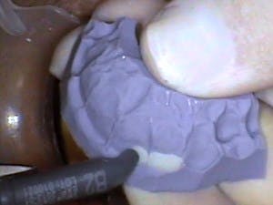
Using a Flexitime® Matrix in Anterior Composite Restorations
Feb 17, 2010
Proof of Principle: human histologic demonstration of socket healing with socket shield and grafting using non-demineralized autologous tooth dentin graft
Apr 24, 2023
The Replacement of Small-Diameter Teeth in the Esthetic Zone Using Narrow-Diameter Implants
Sep 20, 2022
4 Steps to a Predictable Full Arch Rehabilitation
Jun 1, 2021
ATTEND EVENTS WORLDWIDE

Date: 5/16/2024
Location: Modena, Italy
.jpg&w=3840&q=75)