Login
Register
Login
Register


Presented by:
Event Date(s): October 24 - 26, 2024
Location: Chantilly, VA, United States


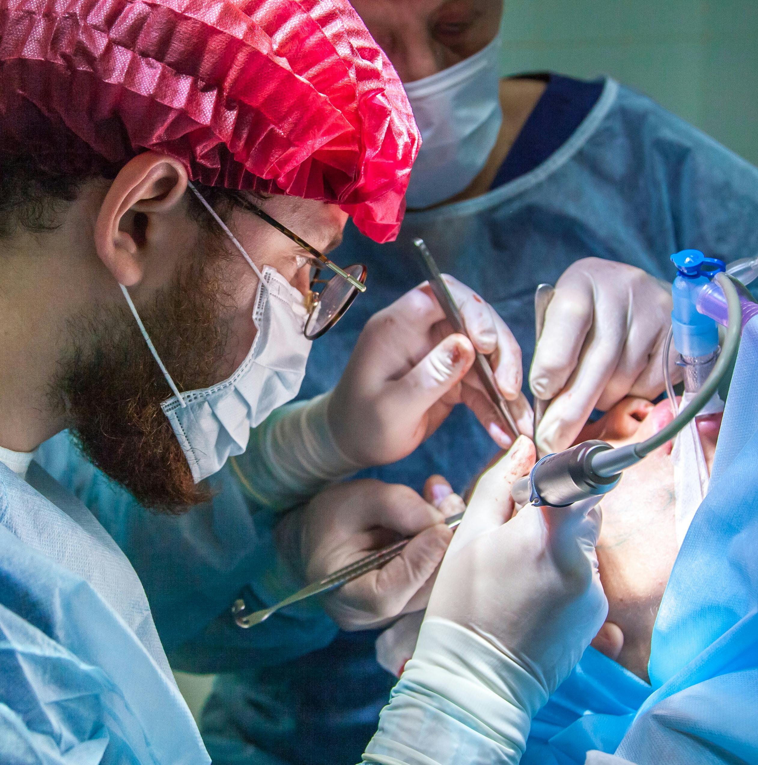
Join us for the 9th edition of the ImplantoDays Congress 6 – 8 June 2024, in the breathtaking setting of Poiana Brașov.
Presented by: Howard Gluckman, BDS, MCHD, PHD, Christian Coachman, Maurice Salama, +5 moreMiguel Stanley, Marcelo Ferrer Balart, Henriette Lerner, Ventseslav Stankov, Roberto Carvalho da Silva
Affiliates: ImplantoDays
Event Date: June 6, 2024
View DetailsUPCOMING WEBINARS
View All Webinars
Webinar Presenter:
Sean Park,Webinar Date: May 15, 2024
CE Hours: 1
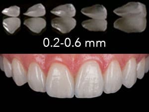
Full monolithic zirconia with external stain technique is one of the popular preference techniques used today. There are many selections of multi-layered zircon...
View Webinar
FROM THE FORUM
View DXP Forum
LIVE EVENTS
View All Events
NEW CONTENT
View All Content
- 5/8/2024
It’s imperative to create a strategy to target patients with a balanced and sensitive approach.
- 5/8/2024
As the saying goes, the best time to review your marketing plan was yesterday. The second best time is today.
OUR CONTINUING DENTAL EDUCATION SPECIALTIES
TRY BEFORE YOU BUY
Using a Flexitime® Matrix in Anterior Composite Restorations
Feb 17, 2010
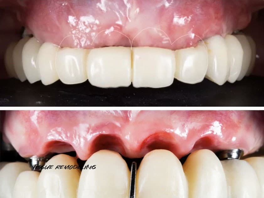
Proof of Principle: human histologic demonstration of socket healing with socket shield and grafting using non-demineralized autologous tooth dentin graft
Apr 24, 2023
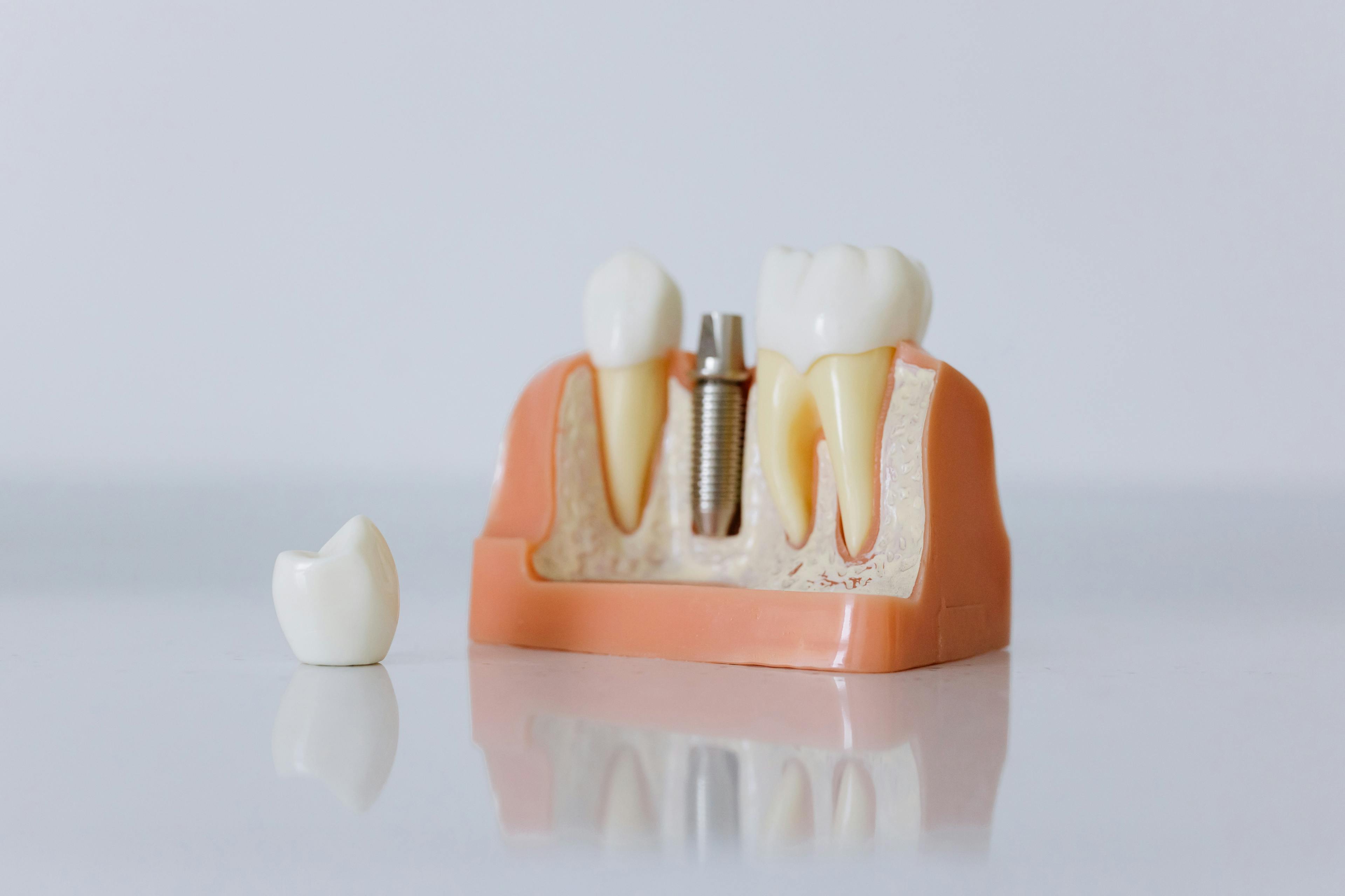
The Replacement of Small-Diameter Teeth in the Esthetic Zone Using Narrow-Diameter Implants
Sep 20, 2022
4 Steps to a Predictable Full Arch Rehabilitation
Jun 1, 2021
ATTEND EVENTS WORLDWIDE

Date: 5/16/2024
Location: Modena, Italy
.jpg&w=3840&q=75)